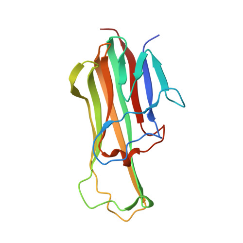
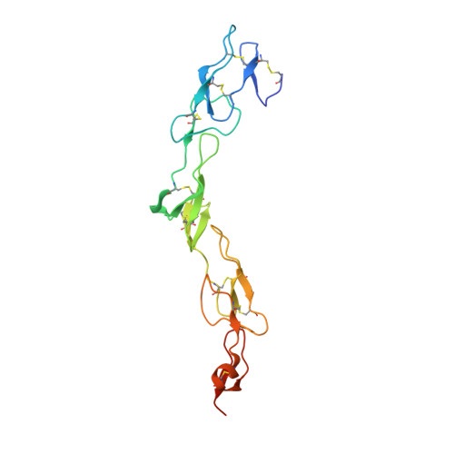
3QBQ

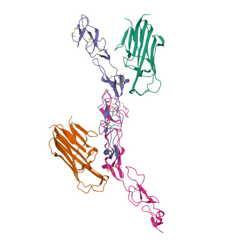
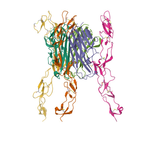
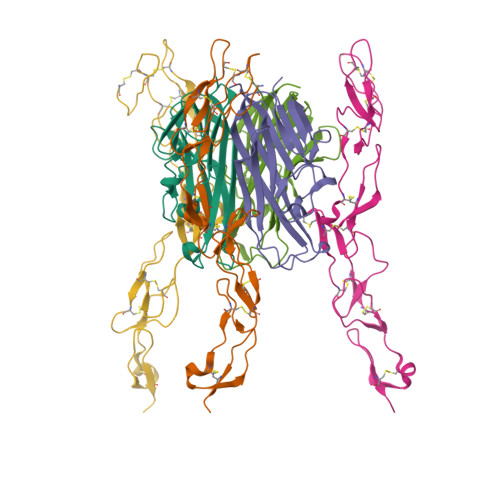
Crystal structure of extracellular domains of mouse RANK-RANKL complex


Experimental Data Snapshot

  • Method: X-RAY DIFFRACTION
  • Resolution: 2.50 Å
  • R-Value Free: 0.249 
  • R-Value Work: 0.227 

Starting Model: experimental
View more details

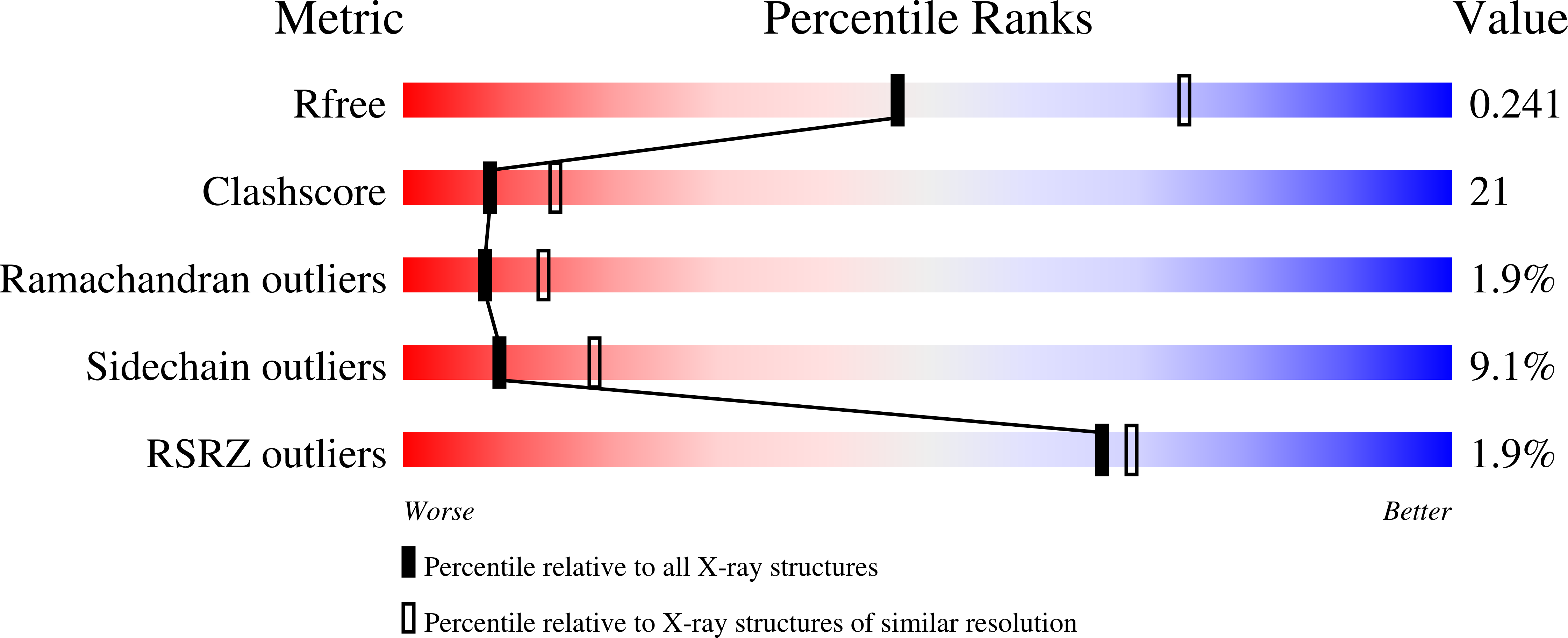
wwPDB Validation   3D Report Full Report


This is version 1.3 of the entry. See complete history


Literature

Structure-based development of a receptor activator of nuclear factor-kappaB ligand (RANKL) inhibitor peptide and molecular basis for osteopetrosis

Ta, H.M.Nguyen, G.T.T.Jin, H.M.Choi, J.K.Park, H.Kim, N.S.Hwang, H.Y.Kim, K.K.

(2010) Proc Natl Acad Sci U S A 107: 20281-20286

  • DOI: https://doi.org/10.1073/pnas.1011686107
  • Primary Citation of Related Structures:  
    3QBQ

  • PubMed Abstract: 

    The receptor activator of nuclear factor-κB (RANK) and its ligand RANKL, which belong to the tumor necrosis factor (TNF) receptor-ligand family, mediate osteoclastogenesis. The crystal structure of the RANKL ectodomain (eRANKL) in complex with the RANK ectodomain (eRANK) combined with biochemical assays of RANK mutants indicated that three RANK loops (Loop1, Loop2, and Loop3) bind to the interface of a trimeric eRANKL. Loop3 is particularly notable in that it is structurally distinctive from other TNF-family receptors and forms extensive contacts with RANKL. The disulfide bond (C125-C127) at the tip of Loop3 is important for determining the unique topology of Loop3, and docking E126 close to RANKL, which was supported by the inability of C127A or E126A mutants of RANK to bind to RANKL. Inhibitory activity of RANK mutants, which contain loops of osteoprotegerin (OPG), a soluble decoy receptor to RANKL, confirmed that OPG shares the similar binding mode with RANK and OPG. Loop3 plays a key role in RANKL binding. Peptide inhibitors designed to mimic Loop3 blocked the RANKL-induced differentiation of osteoclast precursors, suggesting that they could be developed as therapeutic agents for the treatment of osteoporosis and bone-related diseases. Furthermore, some of the RANK mutations associated with autosomal recessive osteopetrosis (ARO) resulted in reduced RANKL-binding activity and failure to induce osteoclastogenesis. These results, together with structural interpretation of eRANK-eRANKL interaction, provided molecular understanding for pathogenesis of ARO.


  • Organizational Affiliation

    Department of Molecular Cell Biology, Samsung Biomedical Research Institute, Sungkyunkwan University School of Medicine, Suwon 440-746, Korea.


Macromolecules
Find similar proteins by:  (by identity cutoff)  |  3D Structure
Entity ID: 1
MoleculeChains Sequence LengthOrganismDetailsImage
Tumor necrosis factor ligand superfamily member 11
A, C
160Mus musculusMutation(s): 0 
Gene Names: Rankl
UniProt
Find proteins for O35235 (Mus musculus)
Explore O35235 
Go to UniProtKB:  O35235
Entity Groups  
Sequence Clusters30% Identity50% Identity70% Identity90% Identity95% Identity100% Identity
UniProt GroupO35235
Sequence Annotations
Expand
  • Reference Sequence
Find similar proteins by:  (by identity cutoff)  |  3D Structure
Entity ID: 2
MoleculeChains Sequence LengthOrganismDetailsImage
Tumor necrosis factor receptor superfamily member 11A
B, D
170Mus musculusMutation(s): 0 
Gene Names: Rank
UniProt
Find proteins for O35305 (Mus musculus)
Explore O35305 
Go to UniProtKB:  O35305
Entity Groups  
Sequence Clusters30% Identity50% Identity70% Identity90% Identity95% Identity100% Identity
UniProt GroupO35305
Sequence Annotations
Expand
  • Reference Sequence
Experimental Data & Validation

Experimental Data

  • Method: X-RAY DIFFRACTION
  • Resolution: 2.50 Å
  • R-Value Free: 0.249 
  • R-Value Work: 0.227 
  • Space Group: P 3
Unit Cell:
Length ( Å )Angle ( ˚ )
a = 120.86α = 90
b = 120.86β = 90
c = 93.425γ = 120
Software Package:
Software NamePurpose
CNSrefinement
PDB_EXTRACTdata extraction
HKL-2000data collection
DENZOdata reduction
SCALEPACKdata scaling

Structure Validation

View Full Validation Report


Entry History 

Deposition Data

Revision History  (Full details and data files)

  • Version 1.0: 2011-03-02
    Type: Initial release
  • Version 1.1: 2011-07-13
    Changes: Version format compliance
  • Version 1.2: 2023-11-01
    Changes: Data collection, Database references, Refinement description
  • Version 1.3: 2024-10-16
    Changes: Structure summary