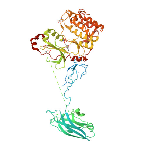
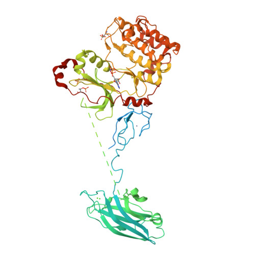
3PFQ

Crystal Structure and Allosteric Activation of Protein Kinase C beta II


Experimental Data Snapshot

  • Method: X-RAY DIFFRACTION
  • Resolution: 4.00 Å
  • R-Value Free: 0.244 
  • R-Value Work: 0.193 
  • R-Value Observed: 0.244 

Starting Models: experimental
View more details

wwPDB Validation   3D Report Full Report


Ligand Structure Quality Assessment 


This is version 1.5 of the entry. See complete history


Literature

Crystal Structure and Allosteric Activation of Protein Kinase C beta II

Leonard, T.A.Rozycki, B.Saidi, L.F.Hummer, G.Hurley, J.H.

(2011) Cell 144: 55-66

  • DOI: https://doi.org/10.1016/j.cell.2010.12.013
  • Primary Citation of Related Structures:  
    3PFQ

  • PubMed Abstract: 

    Protein kinase C (PKC) isozymes are the paradigmatic effectors of lipid signaling. PKCs translocate to cell membranes and are allosterically activated upon binding of the lipid diacylglycerol to their C1A and C1B domains. The crystal structure of full-length protein kinase C βII was determined at 4.0 Å, revealing the conformation of an unexpected intermediate in the activation pathway. Here, the kinase active site is accessible to substrate, yet the conformation of the active site corresponds to a low-activity state because the ATP-binding side chain of Phe629 of the conserved NFD motif is displaced. The C1B domain clamps the NFD helix in a low-activity conformation, which is reversed upon membrane binding. A low-resolution solution structure of the closed conformation of PKCβII was derived from small-angle X-ray scattering. Together, these results show how PKCβII is allosterically regulated in two steps, with the second step defining a novel protein kinase regulatory mechanism.


  • Organizational Affiliation

    Laboratory of Molecular Biology, National Institutes of Health, Bethesda, MD 20892, USA.


Macromolecules
Find similar proteins by:  (by identity cutoff)  |  3D Structure
Entity ID: 1
MoleculeChains Sequence LengthOrganismDetailsImage
Protein kinase C beta type674Rattus norvegicusMutation(s): 3 
Gene Names: PrkcbPkcbPrkcb1
EC: 2.7.11.13
Membrane Entity: Yes 
UniProt
Find proteins for P68403 (Rattus norvegicus)
Explore P68403 
Go to UniProtKB:  P68403
Entity Groups  
Sequence Clusters30% Identity50% Identity70% Identity90% Identity95% Identity100% Identity
UniProt GroupP68403
Sequence Annotations
Expand
  • Reference Sequence
Small Molecules
Modified Residues  2 Unique
IDChains TypeFormula2D DiagramParent
SEP
Query on SEP
A
L-PEPTIDE LINKINGC3 H8 N O6 PSER
TPO
Query on TPO
A
L-PEPTIDE LINKINGC4 H10 N O6 PTHR
Experimental Data & Validation

Experimental Data

  • Method: X-RAY DIFFRACTION
  • Resolution: 4.00 Å
  • R-Value Free: 0.244 
  • R-Value Work: 0.193 
  • R-Value Observed: 0.244 
  • Space Group: P 32 2 1
Unit Cell:
Length ( Å )Angle ( ˚ )
a = 114.27α = 90
b = 114.27β = 90
c = 170.84γ = 120
Software Package:
Software NamePurpose
JBluIce-EPICSdata collection
PHASERphasing
CNSrefinement
MOSFLMdata reduction
SCALAdata scaling

Structure Validation

View Full Validation Report


Ligand Structure Quality Assessment 


Entry History 

Deposition Data

Revision History  (Full details and data files)

  • Version 1.0: 2011-02-02
    Type: Initial release
  • Version 1.1: 2011-07-13
    Changes: Version format compliance
  • Version 1.2: 2017-11-08
    Changes: Refinement description
  • Version 1.3: 2018-04-11
    Changes: Data collection, Database references, Derived calculations, Source and taxonomy, Structure summary
  • Version 1.4: 2023-09-06
    Changes: Data collection, Database references, Derived calculations, Refinement description
  • Version 1.5: 2024-10-16
    Changes: Structure summary