2R03

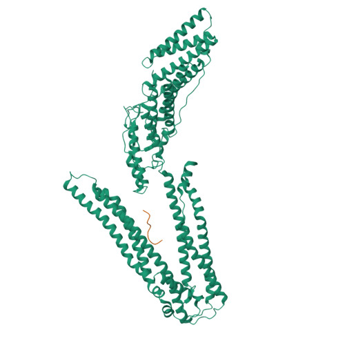
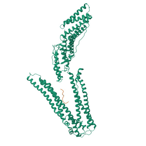
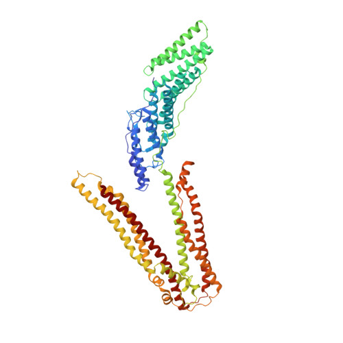
Crystal Structure of ALIX/AIP1 in complex with the YPDL Late Domain


Experimental Data Snapshot

  • Method: X-RAY DIFFRACTION
  • Resolution: 2.59 Å
  • R-Value Free: 0.286 
  • R-Value Work: 0.223 
  • R-Value Observed: 0.226 

wwPDB Validation   3D Report Full Report


This is version 1.4 of the entry. See complete history


Literature

Structural and functional studies of ALIX interactions with YPX(n)L late domains of HIV-1 and EIAV.

Zhai, Q.Fisher, R.D.Chung, H.Y.Myszka, D.G.Sundquist, W.I.Hill, C.P.

(2008) Nat Struct Mol Biol 15: 43-49

  • DOI: https://doi.org/10.1038/nsmb1319
  • Primary Citation of Related Structures:  
    2R02, 2R03, 2R05

  • PubMed Abstract: 

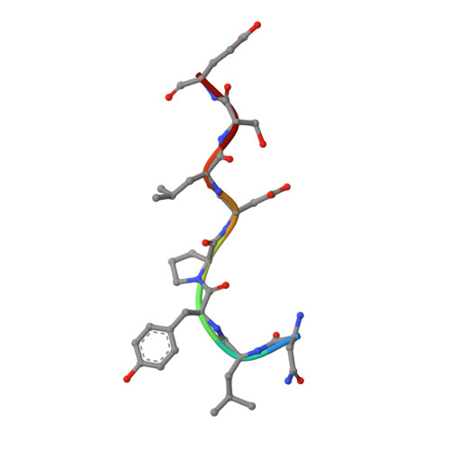
    Retrovirus budding requires short peptide motifs (late domains) located within the viral Gag protein that function by recruiting cellular factors. The YPX(n)L late domains of HIV and other lentiviruses recruit the protein ALIX (also known as AIP1), which also functions in vesicle formation at the multivesicular body and in the abscission stage of cytokinesis. Here, we report the crystal structures of ALIX in complex with the YPX(n)L late domains from HIV-1 and EIAV. The two distinct late domains bind at the same site on the ALIX V domain but adopt different conformations that allow them to make equivalent contacts. Binding studies and functional assays verified the importance of key interface residues and revealed that binding affinities are tuned by context-dependent effects. These results reveal how YPX(n)L late domains recruit ALIX to facilitate virus budding and how ALIX can bind YPX(n)L sequences with both n = 1 and n = 3.


  • Organizational Affiliation

    Department of Biochemistry, University of Utah School of Medicine, Salt Lake City, Utah 84112-5650, USA.


Macromolecules
Find similar proteins by:  (by identity cutoff)  |  3D Structure
Entity ID: 1
MoleculeChains Sequence LengthOrganismDetailsImage
Programmed cell death 6-interacting protein697Homo sapiensMutation(s): 2 
Gene Names: PDCD6IPAIP1ALIXKIAA1375
UniProt & NIH Common Fund Data Resources
Find proteins for Q8WUM4 (Homo sapiens)
Explore Q8WUM4 
Go to UniProtKB:  Q8WUM4
PHAROS:  Q8WUM4
GTEx:  ENSG00000170248 
Entity Groups  
Sequence Clusters30% Identity50% Identity70% Identity90% Identity95% Identity100% Identity
UniProt GroupQ8WUM4
Sequence Annotations
Expand
  • Reference Sequence

Find similar proteins by:  Sequence   |   3D Structure  

Entity ID: 2
MoleculeChains Sequence LengthOrganismDetailsImage
p6-Gag8N/AMutation(s): 0 
UniProt
Find proteins for P69732 (Equine infectious anemia virus (strain Wyoming))
Explore P69732 
Go to UniProtKB:  P69732
Entity Groups  
UniProt GroupP69732
Sequence Annotations
Expand
  • Reference Sequence
Experimental Data & Validation

Experimental Data

  • Method: X-RAY DIFFRACTION
  • Resolution: 2.59 Å
  • R-Value Free: 0.286 
  • R-Value Work: 0.223 
  • R-Value Observed: 0.226 
  • Space Group: C 1 2 1
Unit Cell:
Length ( Å )Angle ( ˚ )
a = 146.383α = 90
b = 98.57β = 107.07
c = 72.799γ = 90
Software Package:
Software NamePurpose
DENZOdata reduction
SCALEPACKdata scaling
REFMACrefinement
PDB_EXTRACTdata extraction
HKL-2000data collection
HKL-2000data reduction

Structure Validation

View Full Validation Report


Entry History 

Deposition Data

Revision History  (Full details and data files)

  • Version 1.0: 2007-12-18
    Type: Initial release
  • Version 1.1: 2011-07-13
    Changes: Version format compliance
  • Version 1.2: 2017-10-25
    Changes: Refinement description
  • Version 1.3: 2021-10-20
    Changes: Database references
  • Version 1.4: 2024-02-21
    Changes: Data collection