2H1K

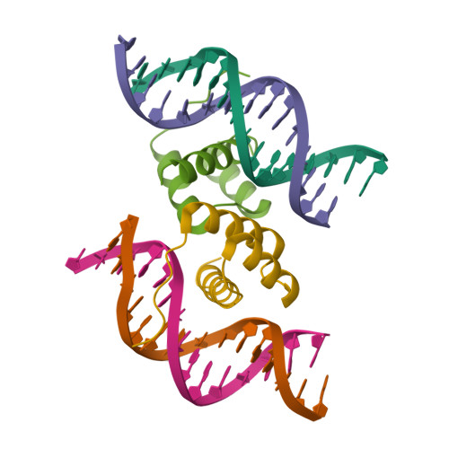
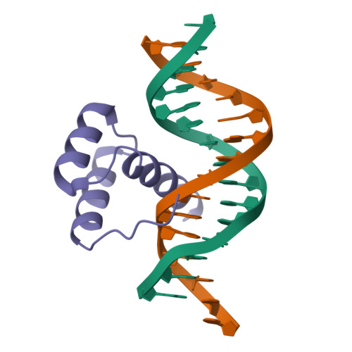
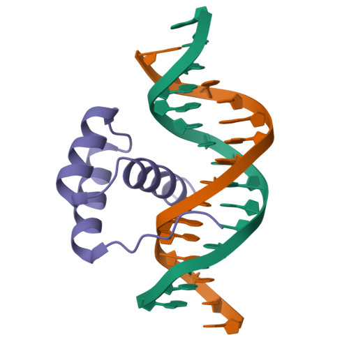
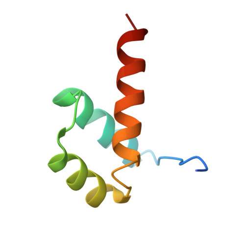
Crystal structure of the Pdx1 homeodomain in complex with DNA


Experimental Data Snapshot

  • Method: X-RAY DIFFRACTION
  • Resolution: 2.42 Å
  • R-Value Free: 0.277 
  • R-Value Work: 0.227 

Starting Model: experimental
View more details

wwPDB Validation   3D Report Full Report


This is version 1.3 of the entry. See complete history


Literature

Structural basis for induced fit mechanisms in DNA recognition by the pdx1 homeodomain

Longo, A.Guanga, G.P.Rose, R.B.

(2007) Biochemistry 46: 2948-2957

  • DOI: https://doi.org/10.1021/bi060969l
  • Primary Citation of Related Structures:  
    2H1K

  • PubMed Abstract: 

    Pancreatic and duodenal homeobox 1 (Pdx1) is a homeodomain transcription factor belonging to the ParaHox family. Pdx1 plays an essential role in pancreatic endocrine and exocrine cell development and maintenance of adult islet beta-cell function. Mutations in the human pdx1 gene are linked to an early onset form of non-insulin-dependent diabetes mellitus, MODY-4. We demonstrate that the homeodomain reproduces the binding specificity of the full-length protein. We report the 2.4 A resolution crystal structure of the homeodomain bound to a target DNA. The two Pdx1/DNA complexes in the asymmetric unit display conformational differences: in the DNA curvature, the orientation of the homeodomain in the major groove, and the order of the N-terminal arm. Comparing the two complexes indicates invariant protein-DNA contacts, and variant contacts that are unique to each binding orientation. An induced fit model is proposed that depends on the DNA conformation and provides a mechanism for nonlocal contributions to binding specificity.


  • Organizational Affiliation

    Department of Molecular and Structural Biochemistry, 128 Polk Hall, North Carolina State University, Raleigh, North Carolina 27695, USA.


Macromolecules

Find similar proteins by:  (by identity cutoff)  |  3D Structure
Entity ID: 3
MoleculeChains Sequence LengthOrganismDetailsImage
Pancreatic and duodenal homeobox 1E [auth A],
F [auth B]
63Mesocricetus auratusMutation(s): 0 
Gene Names: IPF1PDX1
UniProt
Find proteins for P70118 (Mesocricetus auratus)
Explore P70118 
Go to UniProtKB:  P70118
Entity Groups  
Sequence Clusters30% Identity50% Identity70% Identity90% Identity95% Identity100% Identity
UniProt GroupP70118
Sequence Annotations
Expand
  • Reference Sequence

Find similar nucleic acids by:  Sequence   |   3D Structure  

Entity ID: 1
MoleculeChains LengthOrganismImage
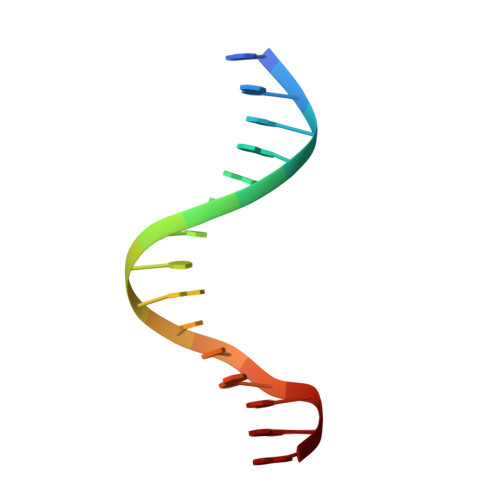
15-mer DNAA [auth C],
C [auth E]
15N/A
Sequence Annotations
Expand
  • Reference Sequence

Find similar nucleic acids by:  Sequence   |   3D Structure  

Entity ID: 2
MoleculeChains LengthOrganismImage
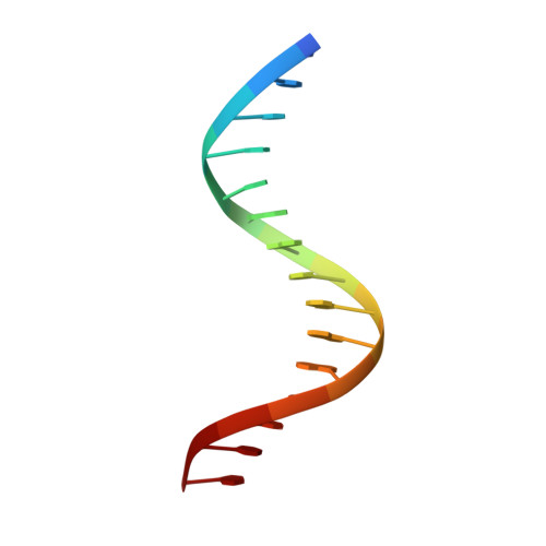
15-mer DNAB [auth D],
D [auth F]
15N/A
Sequence Annotations
Expand
  • Reference Sequence
Experimental Data & Validation

Experimental Data

  • Method: X-RAY DIFFRACTION
  • Resolution: 2.42 Å
  • R-Value Free: 0.277 
  • R-Value Work: 0.227 
  • Space Group: P 21 21 21
Unit Cell:
Length ( Å )Angle ( ˚ )
a = 58.282α = 90
b = 61.928β = 90
c = 96.445γ = 90
Software Package:
Software NamePurpose
ADSCdata collection
MOSFLMdata reduction
CNSrefinement
CNSphasing

Structure Validation

View Full Validation Report


Entry History 

Deposition Data

Revision History  (Full details and data files)

  • Version 1.0: 2007-03-20
    Type: Initial release
  • Version 1.1: 2008-05-01
    Changes: Version format compliance
  • Version 1.2: 2011-07-13
    Changes: Version format compliance
  • Version 1.3: 2023-08-30
    Changes: Data collection, Database references, Refinement description