Cyanide binding to Lucina pectinata hemoglobin I and to sperm whale myoglobin: an x-ray crystallographic study.
Bolognesi, M., Rosano, C., Losso, R., Borassi, A., Rizzi, M., Wittenberg, J.B., Boffi, A., Ascenzi, P.(1999) Biophys J 77: 1093-1099
- PubMed: 10423453
- DOI: https://doi.org/10.1016/S0006-3495(99)76959-6
- Primary Citation of Related Structures:
1B0B, 1EBC, 1EBT - PubMed Abstract:
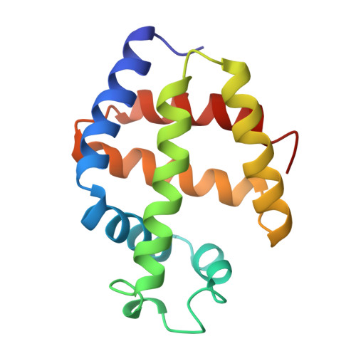
The x-ray crystal structures of the cyanide derivative of Lucina pectinata monomeric hemoglobin I (L. pectinata HbI) and sperm whale (Physeter catodon) myoglobin (Mb), generally taken as reference models for monomeric hemoproteins carrying hydrogen sulfide and oxygen, respectively, have been determined at 1.9 A (R-factor = 0. 184), and 1.8 A (R-factor = 0.181) resolution, respectively, at room temperature (lambda = 1.542 A). Moreover, the x-ray crystal structure of the L. pectinata HbI:cyanide derivative has been studied at 1.4-A resolution (R-factor = 0.118) and 100 K (on a synchrotron source lambda = 0.998 A). At room temperature, the cyanide ligand is roughly parallel to the heme plane of L. pectinata HbI, being located approximately 2.5 A from the iron atom. On the other hand, the crystal structure of the L. pectinata HbI:cyanide derivative at 100 K shows that the diatomic ligand is coordinated to the iron atom in an orientation almost perpendicular to the heme (the Fe-C distance being 1.95 A), adopting a coordination geometry strictly reminescent of that observed in sperm whale Mb, at room temperature. The unusual cyanide distal site orientation observed in L. pectinata HbI, at room temperature, may reflect reduction of the heme Fe(III) atom induced by free radical species during x-ray data collection using Cu Kalpha radiation.
Organizational Affiliation:
Dipartimento di Fisica-INFM, Università di Genova, and Centro Biotecnologie Avanzate-IST, I-16132 Genova, Italy. bolognes@fisica.unige.it