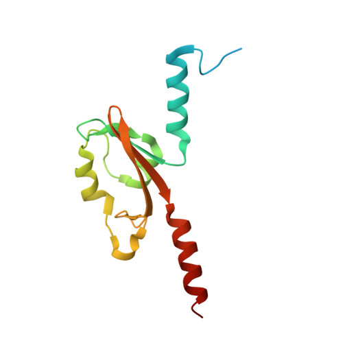
7YRT

Crystal structure of PAS like domain of FlrB, the histidine kinase involved in flagellar synthesis of Vibrio cholerae


Experimental Data Snapshot

  • Method: X-RAY DIFFRACTION
  • Resolution: 2.27 Å
  • R-Value Free: 0.251 
  • R-Value Work: 0.185 
  • R-Value Observed: 0.188 

Starting Model: in silico
View more details

wwPDB Validation   3D Report Full Report


This is version 2.0 of the entry. See complete history


Literature

PAS domain of flagellar histidine kinase FlrB has a unique architecture and binds heme as a sensory ligand in an unconventional fashion.

Mukherjee, P.Agarwal, S.Mallick, S.B.Dasgupta, J.

(2024) Structure 32: 200-216.e5

  • DOI: https://doi.org/10.1016/j.str.2023.11.014
  • Primary Citation of Related Structures:  
    7YRT

  • PubMed Abstract: 

    Phosphorylation of the σ 54 -dependent transcription activator FlrC by the sensor histidine kinase FlrB is essential for flagellar synthesis of Vibrio cholerae. Despite that, the structure, sensory signal, and mechanistic basis of function of FlrB were elusive. Here, we report the crystal structure of the sensory PAS domain of FlrB in its functional dimeric state that exhibits a unique architecture. Series of biochemical/biophysical experiments on different constructs and mutants established that heme binds hydrophobically as sensory ligand in the shallow ligand-binding cleft of FlrB-PAS without axial coordination. Intriguingly, ATP binding to the C-terminal ATP-binding (CA) domain assists PAS domain to bind heme, vis-à-vis, heme binding to the PAS facilitates ATP binding to the CA domain. We hypothesize that synergistic binding of heme and ATP triggers conformational signaling in FlrB, leading to downstream flagellar gene transcription. Enhanced swimming motility of V. cholerae with increased heme uptake supports this proposition.


  • Organizational Affiliation

    Department of Biotechnology, St. Xavier's College (Autonomous), 30 Mother Teresa Sarani, Kolkata 700016, India.


Macromolecules
Find similar proteins by:  (by identity cutoff)  |  3D Structure
Entity ID: 1
MoleculeChains Sequence LengthOrganismDetailsImage
histidine kinase
A, B, C, D, E
A, B, C, D, E, F, G, H, I, J, K, L
138Vibrio cholerae O395Mutation(s): 0 
Gene Names: flrBVC0395_A1720
EC: 2.7.13.3
UniProt
Find proteins for H9L4P6 (Vibrio cholerae serotype O1 (strain ATCC 39315 / El Tor Inaba N16961))
Explore H9L4P6 
Go to UniProtKB:  H9L4P6
Entity Groups  
Sequence Clusters30% Identity50% Identity70% Identity90% Identity95% Identity100% Identity
UniProt GroupH9L4P6
Sequence Annotations
Expand
  • Reference Sequence
Experimental Data & Validation

Experimental Data

  • Method: X-RAY DIFFRACTION
  • Resolution: 2.27 Å
  • R-Value Free: 0.251 
  • R-Value Work: 0.185 
  • R-Value Observed: 0.188 
  • Space Group: C 1 2 1
Unit Cell:
Length ( Å )Angle ( ˚ )
a = 212.644α = 90
b = 49.556β = 92.53
c = 146.83γ = 90
Software Package:
Software NamePurpose
PHENIXrefinement
XDSdata reduction
Aimlessdata scaling
PHASERphasing

Structure Validation

View Full Validation Report


Entry History & Funding Information

Deposition Data


Funding OrganizationLocationGrant Number
Department of Science & Technology (DST, India)India--

Revision History  (Full details and data files)

  • Version 1.0: 2022-10-05
    Type: Initial release
  • Version 1.1: 2024-04-03
    Changes: Data collection, Refinement description
  • Version 2.0: 2024-05-01
    Changes: Advisory, Atomic model, Database references, Derived calculations, Polymer sequence, Source and taxonomy, Structure summary