5F74

Crystal structure of ChREBP:14-3-3 complex bound with AMP


Experimental Data Snapshot

  • Method: X-RAY DIFFRACTION
  • Resolution: 2.35 Å
  • R-Value Free: 0.255 
  • R-Value Work: 0.188 
  • R-Value Observed: 0.191 

Starting Model: experimental
View more details

wwPDB Validation   3D Report Full Report


Ligand Structure Quality Assessment 


This is version 1.3 of the entry. See complete history


Literature

Metabolite Regulation of Nuclear Localization of Carbohydrate-response Element-binding Protein (ChREBP): ROLE OF AMP AS AN ALLOSTERIC INHIBITOR.

Sato, S.Jung, H.Nakagawa, T.Pawlosky, R.Takeshima, T.Lee, W.R.Sakiyama, H.Laxman, S.Wynn, R.M.Tu, B.P.MacMillan, J.B.De Brabander, J.K.Veech, R.L.Uyeda, K.

(2016) J Biol Chem 291: 10515-10527

  • DOI: https://doi.org/10.1074/jbc.M115.708982
  • Primary Citation of Related Structures:  
    5F74

  • PubMed Abstract: 

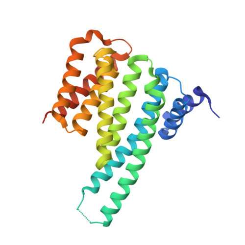
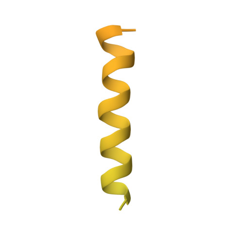
    The carbohydrate-response element-binding protein (ChREBP) is a glucose-responsive transcription factor that plays an essential role in converting excess carbohydrate to fat storage in the liver. In response to glucose levels, ChREBP is regulated by nuclear/cytosol trafficking via interaction with 14-3-3 proteins, CRM-1 (exportin-1 or XPO-1), or importins. Nuclear localization of ChREBP was rapidly inhibited when incubated in branched-chain α-ketoacids, saturated and unsaturated fatty acids, or 5-aminoimidazole-4-carboxamide ribonucleotide. Here, we discovered that protein-free extracts of high fat-fed livers contained, in addition to ketone bodies, a new metabolite, identified as AMP, which specifically activates the interaction between ChREBP and 14-3-3. The crystal structure showed that AMP binds directly to the N terminus of ChREBP-α2 helix. Our results suggest that AMP inhibits the nuclear localization of ChREBP through an allosteric activation of ChREBP/14-3-3 interactions and not by activation of AMPK. AMP and ketone bodies together can therefore inhibit lipogenesis by restricting localization of ChREBP to the cytoplasm during periods of ketosis.


  • Organizational Affiliation

    From the Department of Biochemistry, University of Texas Southwestern Medical Center at Dallas, Dallas, Texas 75390.


Macromolecules
Find similar proteins by:  (by identity cutoff)  |  3D Structure
Entity ID: 1
MoleculeChains Sequence LengthOrganismDetailsImage
14-3-3 protein beta/alpha246Mus musculusMutation(s): 0 
Gene Names: Ywhab
UniProt & NIH Common Fund Data Resources
Find proteins for Q9CQV8 (Mus musculus)
Explore Q9CQV8 
Go to UniProtKB:  Q9CQV8
IMPC:  MGI:1891917
Entity Groups  
Sequence Clusters30% Identity50% Identity70% Identity90% Identity95% Identity100% Identity
UniProt GroupQ9CQV8
Sequence Annotations
Expand
  • Reference Sequence
Find similar proteins by:  (by identity cutoff)  |  3D Structure
Entity ID: 2
MoleculeChains Sequence LengthOrganismDetailsImage
Carbohydrate-responsive element-binding protein196Rattus norvegicusMutation(s): 0 
Gene Names: MlxiplWbscr14
UniProt
Find proteins for Q8VIP2 (Rattus norvegicus)
Explore Q8VIP2 
Go to UniProtKB:  Q8VIP2
Entity Groups  
Sequence Clusters30% Identity50% Identity70% Identity90% Identity95% Identity100% Identity
UniProt GroupQ8VIP2
Sequence Annotations
Expand
  • Reference Sequence
Small Molecules
Ligands 1 Unique
IDChains Name / Formula / InChI Key2D Diagram3D Interactions
AMP
Query on AMP

Download Ideal Coordinates CCD File 
C [auth A]ADENOSINE MONOPHOSPHATE
C10 H14 N5 O7 P
UDMBCSSLTHHNCD-KQYNXXCUSA-N
Experimental Data & Validation

Experimental Data

  • Method: X-RAY DIFFRACTION
  • Resolution: 2.35 Å
  • R-Value Free: 0.255 
  • R-Value Work: 0.188 
  • R-Value Observed: 0.191 
  • Space Group: P 42 2 2
Unit Cell:
Length ( Å )Angle ( ˚ )
a = 115.62α = 90
b = 115.62β = 90
c = 59.372γ = 90
Software Package:
Software NamePurpose
PHENIXrefinement
HKL-3000data reduction
HKL-3000data scaling
MOLREPphasing

Structure Validation

View Full Validation Report


Ligand Structure Quality Assessment 


Entry History 

Deposition Data

Revision History  (Full details and data files)

  • Version 1.0: 2016-03-23
    Type: Initial release
  • Version 1.1: 2016-03-30
    Changes: Database references
  • Version 1.2: 2016-06-01
    Changes: Database references
  • Version 1.3: 2023-09-27
    Changes: Data collection, Database references, Derived calculations, Refinement description