8S4D | pdb_00008s4d

Crystal structure of a peptidergic GPCR in complex with a small synthetic G protein-biased agonist


Experimental Data Snapshot

  • Method: X-RAY DIFFRACTION
  • Resolution: 2.58 Å
  • R-Value Free: 
    0.275 (Depositor), 0.223 (DCC) 
  • R-Value Work: 
    0.232 (Depositor), 0.234 (DCC) 
  • R-Value Observed: 
    0.234 (Depositor) 

Starting Model: experimental
View more details

wwPDB Validation   3D Report Full Report


Ligand Structure Quality Assessment 

Created with Raphaël 2.3.0Worse 01 BetterLigand structure goodness of fit to experimental dataBest fitted A1D5NClick on this verticalbar to view details

This is version 1.1 of the entry. See complete history


Literature

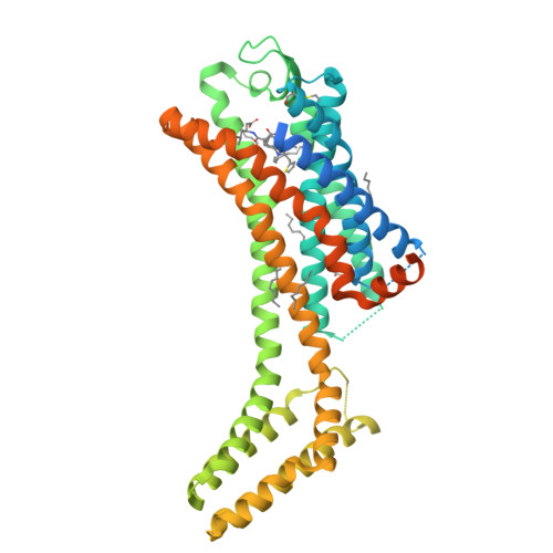
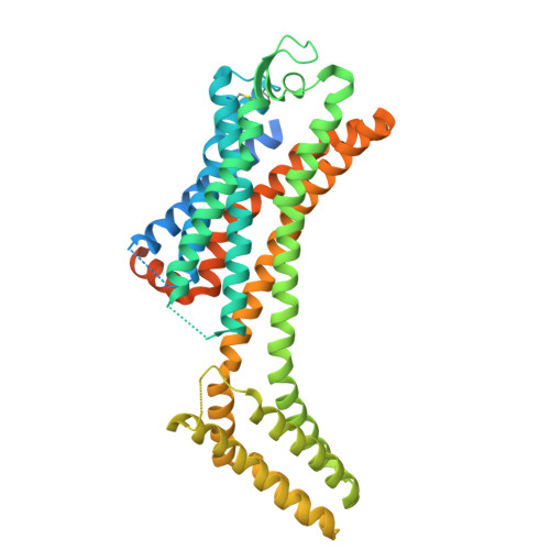
Structural and functional determination of peptide versus small molecule ligand binding at the apelin receptor.

Williams, T.L.Verdon, G.Kuc, R.E.Currinn, H.Bender, B.Solcan, N.Schlenker, O.Macrae, R.G.C.Brown, J.Schutz, M.Zhukov, A.Sinha, S.de Graaf, C.Graf, S.Maguire, J.J.Brown, A.J.H.Davenport, A.P.

(2024) Nat Commun 15: 10714-10714

  • DOI: https://doi.org/10.1038/s41467-024-55381-w
  • Primary Citation of Related Structures:  
    8S4D

  • PubMed Abstract: 

    We describe a structural and functional study of the G protein-coupled apelin receptor, which binds two endogenous peptide ligands, apelin and Elabela/Toddler (ELA), to regulate cardiovascular development and function. Characterisation of naturally occurring apelin receptor variants from the UK Genomics England 100,000 Genomes Project, and AlphaFold2 modelling, identifies T89 2.64 as important in the ELA binding site, and R168 4.64 as forming extensive interactions with the C-termini of both peptides. Base editing to introduce an R/H168 4.64 variant into human stem cell-derived cardiomyocytes demonstrates that this residue is critical for receptor binding and function. Additionally, we present an apelin receptor crystal structure bound to the G protein-biased, small molecule agonist, CMF-019, which reveals a deeper binding mode versus the endogenous peptides at lipophilic pockets between transmembrane helices associated with GPCR activation. Overall, the data provide proof-of-principle for using genetic variation to identify key sites regulating receptor-ligand engagement.


  • Organizational Affiliation

    Experimental Medicine & Immunotherapeutics, University of Cambridge, Cambridge, UK.


Macromolecules
Find similar proteins by:  (by identity cutoff)  |  3D Structure
Entity ID: 1
MoleculeChains Sequence LengthOrganismDetailsImage
Apelin receptor,Soluble cytochrome b562497Homo sapiensEscherichia coli O139:H28 str. E24377AMutation(s): 7 
Gene Names: APLNRAGTRL1APJcybCEcE24377A_4806
UniProt & NIH Common Fund Data Resources
Find proteins for P0ABE7 (Escherichia coli)
Explore P0ABE7 
Go to UniProtKB:  P0ABE7
Find proteins for P35414 (Homo sapiens)
Explore P35414 
Go to UniProtKB:  P35414
PHAROS:  P35414
GTEx:  ENSG00000134817 
Entity Groups  
Sequence Clusters30% Identity50% Identity70% Identity90% Identity95% Identity100% Identity
UniProt GroupsP0ABE7P35414
Sequence Annotations
Expand
  • Reference Sequence
Small Molecules
Ligands 3 Unique
IDChains Name / Formula / InChI Key2D Diagram3D Interactions
A1D5N (Subject of Investigation/LOI)
Query on A1D5N

Download Ideal Coordinates CCD File 
B [auth A](3~{S})-5-methyl-3-[[1-pentan-3-yl-2-(thiophen-2-ylmethyl)benzimidazol-5-yl]carbonylamino]hexanoic acid
C25 H33 N3 O3 S
VCQKKZXFASLXAH-SFHVURJKSA-N
OLA
Query on OLA

Download Ideal Coordinates CCD File 
C [auth A]
D [auth A]
E [auth A]
F [auth A]
G [auth A]
OLEIC ACID
C18 H34 O2
ZQPPMHVWECSIRJ-KTKRTIGZSA-N
1PE
Query on 1PE

Download Ideal Coordinates CCD File 
I [auth A]PENTAETHYLENE GLYCOL
C10 H22 O6
JLFNLZLINWHATN-UHFFFAOYSA-N
Experimental Data & Validation

Experimental Data

  • Method: X-RAY DIFFRACTION
  • Resolution: 2.58 Å
  • R-Value Free:  0.275 (Depositor), 0.223 (DCC) 
  • R-Value Work:  0.232 (Depositor), 0.234 (DCC) 
  • R-Value Observed: 0.234 (Depositor) 
Space Group: C 2 2 21
Unit Cell:
Length ( Å )Angle ( ˚ )
a = 65.823α = 90
b = 153.948β = 90
c = 111.646γ = 90
Software Package:
Software NamePurpose
BUSTERrefinement
Cootmodel building
GDAdata collection
XDSdata reduction
STARANISOdata scaling
PHASERphasing

Structure Validation

View Full Validation Report


Ligand Structure Quality Assessment 

Created with Raphaël 2.3.0Worse 01 BetterLigand structure goodness of fit to experimental dataBest fitted A1D5NClick on this verticalbar to view details

Entry History & Funding Information

Deposition Data


Funding OrganizationLocationGrant Number
Other privateUnited KingdomRG86507

Revision History  (Full details and data files)

  • Version 1.0: 2024-12-04
    Type: Initial release
  • Version 1.1: 2025-01-22
    Changes: Database references