6PII | pdb_00006pii

The evolving story of AtzT, a periplasmic binding protein


Experimental Data Snapshot

  • Method: X-RAY DIFFRACTION
  • Resolution: 1.87 Å
  • R-Value Free: 
    0.183 (Depositor), 0.190 (DCC) 
  • R-Value Work: 
    0.155 (Depositor), 0.170 (DCC) 
  • R-Value Observed: 
    0.156 (Depositor) 

wwPDB Validation   3D Report Full Report


Ligand Structure Quality Assessment 

Created with Raphaël 2.3.0Worse 01 BetterLigand structure goodness of fit to experimental dataBest fitted GUNClick on this verticalbar to view details

This is version 1.2 of the entry. See complete history


Literature

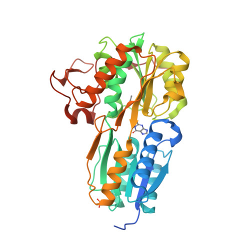
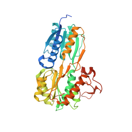
The evolving story of AtzT, a periplasmic binding protein.

Dennis, M.L.Esquirol, L.Nebl, T.Newman, J.Scott, C.Peat, T.S.

(2019) Acta Crystallogr D Struct Biol 75: 995-1002

  • DOI: https://doi.org/10.1107/S2059798319013883
  • Primary Citation of Related Structures:  
    6PI5, 6PI6, 6PII

  • PubMed Abstract: 

    Atrazine is an s-triazine-based herbicide that is used in many countries around the world in many millions of tons per year. A small number of organisms, such as Pseudomonas sp. strain ADP, have evolved to use this modified s-triazine as a food source, and the various genes required to metabolize atrazine can be found on a single plasmid. The atomic structures of seven of the eight proteins involved in the breakdown of atrazine by Pseudomonas sp. strain ADP have been determined by X-ray crystallography, but the structures of the proteins required by the cell to import atrazine for use as an energy source are still lacking. The structure of AtzT, a periplasmic binding protein that may be involved in the transport of a derivative of atrazine, 2-hydroxyatrazine, into the cell for mineralization, has now been determined. The structure was determined by SAD phasing using an ethylmercury phosphate derivative that diffracted X-rays to beyond 1.9 Å resolution. `Native' (guanine-bound) and 2-hydroxyatrazine-bound structures were also determined to high resolution (1.67 and 1.65 Å, respectively), showing that 2-hydroxyatrazine binds in a similar way to the purportedly native ligand. Structural similarities led to the belief that it may be possible to evolve AtzT from a purine-binding protein to a protein that can bind and detect atrazine in the environment.


  • Organizational Affiliation

    Biomedical Manufacturing Program, CSIRO, 343 Royal Parade, Parkville, VIC 3052, Australia.


Macromolecules
Find similar proteins by:  (by identity cutoff)  |  3D Structure
Entity ID: 1
MoleculeChains Sequence LengthOrganismDetailsImage
Atrazine periplasmic binding protein
A, B, C, D
355Pseudomonas sp. ADPMutation(s): 0 
Gene Names: orf97AOX63_31690
UniProt
Find proteins for Q936X6 (Pseudomonas sp. (strain ADP))
Explore Q936X6 
Go to UniProtKB:  Q936X6
Entity Groups  
Sequence Clusters30% Identity50% Identity70% Identity90% Identity95% Identity100% Identity
UniProt GroupQ936X6
Sequence Annotations
Expand
  • Reference Sequence
Small Molecules
Ligands 4 Unique
IDChains Name / Formula / InChI Key2D Diagram3D Interactions
EMC
Query on EMC

Download Ideal Coordinates CCD File 
F [auth A],
I [auth B],
L [auth C],
O [auth D]
ETHYL MERCURY ION
C2 H5 Hg
MJOUBOKSWBMNGQ-UHFFFAOYSA-N
GUN (Subject of Investigation/LOI)
Query on GUN

Download Ideal Coordinates CCD File 
E [auth A],
H [auth B],
K [auth C],
N [auth D]
GUANINE
C5 H5 N5 O
UYTPUPDQBNUYGX-UHFFFAOYSA-N
PO4
Query on PO4

Download Ideal Coordinates CCD File 
G [auth A],
J [auth B]
PHOSPHATE ION
O4 P
NBIIXXVUZAFLBC-UHFFFAOYSA-K
CA
Query on CA

Download Ideal Coordinates CCD File 
M [auth C]CALCIUM ION
Ca
BHPQYMZQTOCNFJ-UHFFFAOYSA-N
Experimental Data & Validation

Experimental Data

  • Method: X-RAY DIFFRACTION
  • Resolution: 1.87 Å
  • R-Value Free:  0.183 (Depositor), 0.190 (DCC) 
  • R-Value Work:  0.155 (Depositor), 0.170 (DCC) 
  • R-Value Observed: 0.156 (Depositor) 
Space Group: P 1 21 1
Unit Cell:
Length ( Å )Angle ( ˚ )
a = 98.92α = 90
b = 63.05β = 93.49
c = 120.436γ = 90
Software Package:
Software NamePurpose
REFMACrefinement
ADDREFdata reduction
Aimlessdata scaling
CRANK2phasing

Structure Validation

View Full Validation Report


Ligand Structure Quality Assessment 

Created with Raphaël 2.3.0Worse 01 BetterLigand structure goodness of fit to experimental dataBest fitted GUNClick on this verticalbar to view details

Entry History 

Deposition Data

Revision History  (Full details and data files)

  • Version 1.0: 2019-11-13
    Type: Initial release
  • Version 1.1: 2019-12-25
    Changes: Database references
  • Version 1.2: 2024-03-13
    Changes: Data collection, Database references, Derived calculations, Refinement description