4POJ | pdb_00004poj

Crystal structure of human Retinoid X Receptor alpha-ligand binding domain complex with 7-methyl UAB30 and the coactivator peptide GRIP-1


Experimental Data Snapshot

  • Method: X-RAY DIFFRACTION
  • Resolution: 2.00 Å
  • R-Value Free: 
    0.261 (Depositor), 0.250 (DCC) 
  • R-Value Work: 
    0.218 (Depositor), 0.220 (DCC) 
  • R-Value Observed: 
    0.218 (Depositor) 

Starting Model: experimental
View more details

wwPDB Validation   3D Report Full Report


Ligand Structure Quality Assessment 

Created with Raphaël 2.3.0Worse 01 BetterLigand structure goodness of fit to experimental dataBest fitted 2VPClick on this verticalbar to view details

This is version 1.3 of the entry. See complete history


Literature

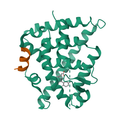
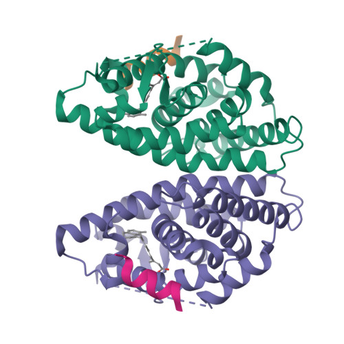
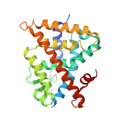
Methyl substitution of a rexinoid agonist improves potency and reveals site of lipid toxicity.

Atigadda, V.R.Xia, G.Desphande, A.Boerma, L.J.Lobo-Ruppert, S.Grubbs, C.J.Smith, C.D.Brouillette, W.J.Muccio, D.D.

(2014) J Med Chem 57: 5370-5380

  • DOI: https://doi.org/10.1021/jm5004792
  • Primary Citation of Related Structures:  
    4POH, 4POJ, 4PP3, 4PP5

  • PubMed Abstract: 

    (2E,4E,6Z,8E)-8-(3',4'-Dihydro-1'(2'H)-naphthalen-1'-ylidene)-3,7-dimethyl-2,4,6-octatrienoic acid, 9cUAB30, is a selective rexinoid that displays substantial chemopreventive capacity with little toxicity. 4-Methyl-UAB30, an analogue of 9cUAB30, is a potent RXR agonist but caused increased lipid biosynthesis unlike 9cUAB30. To evaluate how methyl substitution influenced potency and lipid biosynthesis, we synthesized four 9cUAB30 homologues with methyl substitutions at the 5-, 6-, 7-, or 8-position of the tetralone ring. The syntheses and biological evaluations of these new analogues are reported here along with the X-ray crystal structures of each homologue bound to the ligand binding domain of hRXRα. We demonstrate that each homologue of 9cUAB30 is a more potent agonist, but only the 7-methyl-9cUAB30 caused severe hyperlipidemia in rats. On the basis of the X-ray crystal structures of these new rexinoids and bexarotene (Targretin) bound to hRXRα-LBD, we reveal that each rexinoid, which induced hyperlipidemia, had methyl groups that interacted with helix 7 residues of the LBD.


  • Organizational Affiliation

    Departments of †Chemistry, ‡Biochemistry and Molecular Genetics, §Medicine, and ∥Vision Sciences, University of Alabama at Birmingham , Birmingham, Alabama 35294, United States.


Macromolecules
Find similar proteins by:  (by identity cutoff)  |  3D Structure
Entity ID: 1
MoleculeChains Sequence LengthOrganismDetailsImage
Retinoic acid receptor RXR-alpha231Homo sapiensMutation(s): 0 
Gene Names: NR2B1RXRARXRA NR2B1
UniProt & NIH Common Fund Data Resources
Find proteins for P19793 (Homo sapiens)
Explore P19793 
Go to UniProtKB:  P19793
PHAROS:  P19793
GTEx:  ENSG00000186350 
Entity Groups  
Sequence Clusters30% Identity50% Identity70% Identity90% Identity95% Identity100% Identity
UniProt GroupP19793
Sequence Annotations
Expand
  • Reference Sequence

Find similar proteins by:  Sequence   |   3D Structure  

Entity ID: 2
MoleculeChains Sequence LengthOrganismDetailsImage
Nuclear receptor coactivator 213Homo sapiensMutation(s): 0 
UniProt & NIH Common Fund Data Resources
Find proteins for Q15596 (Homo sapiens)
Explore Q15596 
Go to UniProtKB:  Q15596
PHAROS:  Q15596
GTEx:  ENSG00000140396 
Entity Groups  
Sequence Clusters30% Identity50% Identity70% Identity90% Identity95% Identity100% Identity
UniProt GroupQ15596
Sequence Annotations
Expand
  • Reference Sequence
Small Molecules
Ligands 1 Unique
IDChains Name / Formula / InChI Key2D Diagram3D Interactions
2VP
Query on 2VP

Download Ideal Coordinates CCD File 
C [auth A](2E,4E,6Z,8E)-3,7-dimethyl-8-(7-methyl-3,4-dihydronaphthalen-1(2H)-ylidene)octa-2,4,6-trienoic acid
C21 H24 O2
TZMKYYXLMOFFMM-NDMWHMLYSA-N
Binding Affinity Annotations 
IDSourceBinding Affinity
2VP BindingDB:  4POJ Kd: min: 8, max: 1860 (nM) from 2 assay(s)
EC50: min: 160, max: 1000 (nM) from 2 assay(s)
Experimental Data & Validation

Experimental Data

  • Method: X-RAY DIFFRACTION
  • Resolution: 2.00 Å
  • R-Value Free:  0.261 (Depositor), 0.250 (DCC) 
  • R-Value Work:  0.218 (Depositor), 0.220 (DCC) 
  • R-Value Observed: 0.218 (Depositor) 
Space Group: P 43 21 2
Unit Cell:
Length ( Å )Angle ( ˚ )
a = 65.88α = 90
b = 65.88β = 90
c = 112.01γ = 90
Software Package:
Software NamePurpose
HKL-2000data collection
CNSrefinement
d*TREKdata reduction
d*TREKdata scaling
CNSphasing

Structure Validation

View Full Validation Report


Ligand Structure Quality Assessment 

Created with Raphaël 2.3.0Worse 01 BetterLigand structure goodness of fit to experimental dataBest fitted 2VPClick on this verticalbar to view details

Entry History 

Deposition Data

Revision History  (Full details and data files)

  • Version 1.0: 2014-06-18
    Type: Initial release
  • Version 1.1: 2014-07-23
    Changes: Database references
  • Version 1.2: 2022-08-17
    Changes: Database references, Derived calculations
  • Version 1.3: 2023-09-20
    Changes: Data collection, Refinement description