4OM8 | pdb_00004om8

Crystal structure of 5-formly-3-hydroxy-2-methylpyridine 4-carboxylic acid (FHMPC) 5-dehydrogenase, an NAD+ dependent dismutase.


Experimental Data Snapshot

  • Method: X-RAY DIFFRACTION
  • Resolution: 1.55 Å
  • R-Value Free: 
    0.188 (Depositor), 0.191 (DCC) 
  • R-Value Work: 
    0.155 (Depositor), 0.161 (DCC) 
  • R-Value Observed: 
    0.157 (Depositor) 

Starting Model: experimental
View more details

wwPDB Validation   3D Report Full Report


Ligand Structure Quality Assessment 

Created with Raphaël 2.3.0Worse 01 BetterLigand structure goodness of fit to experimental dataBest fitted NADClick on this verticalbar to view details

This is version 2.0 of the entry. See complete history


Literature

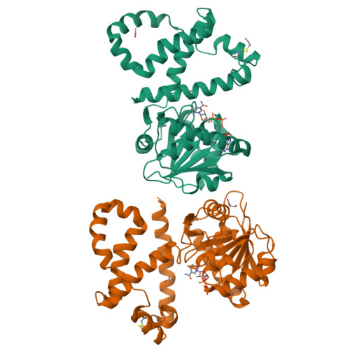
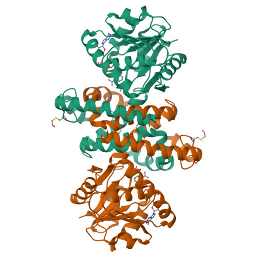
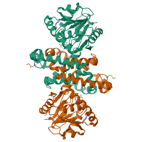
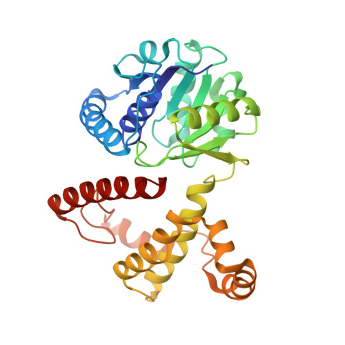
Crystal structure of 5-formyl-3-hydroxy-2-methylpyridine 4-carboxylic acid 5-dehydrogenase, an NAD(+)-dependent dismutase from Mesorhizobium loti

Mugo, A.N.Kobayashi, J.Mikami, B.Yoshikane, Y.Yagi, T.Ohnishi, K.

(2015) Biochem Biophys Res Commun 456: 35-40

  • DOI: https://doi.org/10.1016/j.bbrc.2014.11.028
  • Primary Citation of Related Structures:  
    4OM8

  • PubMed Abstract: 

    5-Formyl-3-hydroxy-2-methylpyridine 4-carboxylic acid 5-dehydrogenase (FHMPCDH) from Mesorhizobium loti is the fifth enzyme in degradation pathway I for pyridoxine. The enzyme catalyzes a dismutation reaction: the oxidation of 5-formyl-3-hydroxy-2-methylpyridine 4-carboxylic acid (FHMPC) to 3-hydroxy-2-methylpyridine 4,5-dicarboxylic acid with NAD(+) and reduction of FHMPC to 4-pyridoxic acid with NADH. FHMPCDH belongs to the l-3-hydroxyacyl-CoA dehydrogenase (HAD) family. The crystal structure was determined by molecular replacement and refined to a resolution of 1.55Å (R-factor of 16.4%, Rfree=19.4%). There were two monomers in the asymmetric unit. The overall structure of the monomer consisted of N- and C-terminal domains connected by a short linker loop. The monomer was similar to members of the HAD family (RMSD=1.9Å). The active site was located between the domains and highly conserved to that of human heart l-3-hydroxyacyl-CoA dehydrogenase (HhHAD). His-Glu catalytic dyad, a serine and two asparagine residues of HhHAD were conserved. Ser116, His137 and Glu149 in FHMPCDH are connected by a hydrogen bonding network forming a catalytic triad. The functions of the active site residues in the reaction mechanism are discussed.


  • Organizational Affiliation

    Research Institute of Molecular Genetics, Kochi University, Nankoku, Kochi 783-8502, Japan.


Macromolecules
Find similar proteins by:  (by identity cutoff)  |  3D Structure
Entity ID: 1
MoleculeChains Sequence LengthOrganismDetailsImage
5-formyl-3-hydroxy-2-methylpyridine 4-carboxylate 5-dehydrogenase
A, B
309Mesorhizobium japonicum MAFF 303099Mutation(s): 0 
Gene Names: fhmpcd1mlr6793
EC: 1.2.1.100
UniProt
Find proteins for Q988C8 (Mesorhizobium japonicum (strain LMG 29417 / CECT 9101 / MAFF 303099))
Explore Q988C8 
Go to UniProtKB:  Q988C8
Entity Groups  
Sequence Clusters30% Identity50% Identity70% Identity90% Identity95% Identity100% Identity
UniProt GroupQ988C8
Sequence Annotations
Expand
  • Reference Sequence
Small Molecules
Modified Residues  1 Unique
IDChains TypeFormula2D DiagramParent
CME
Query on CME
A, B
L-PEPTIDE LINKINGC5 H11 N O3 S2CYS
Experimental Data & Validation

Experimental Data

  • Method: X-RAY DIFFRACTION
  • Resolution: 1.55 Å
  • R-Value Free:  0.188 (Depositor), 0.191 (DCC) 
  • R-Value Work:  0.155 (Depositor), 0.161 (DCC) 
  • R-Value Observed: 0.157 (Depositor) 
Space Group: C 1 2 1
Unit Cell:
Length ( Å )Angle ( ˚ )
a = 122.805α = 90
b = 44.212β = 116.35
c = 109.343γ = 90
Software Package:
Software NamePurpose
REFMACrefinement
PDB_EXTRACTdata extraction
HKL-2000data collection
HKL-2000data reduction
HKL-2000data scaling
MOLREPphasing

Structure Validation

View Full Validation Report


Ligand Structure Quality Assessment 

Created with Raphaël 2.3.0Worse 01 BetterLigand structure goodness of fit to experimental dataBest fitted NADClick on this verticalbar to view details

Entry History 

Deposition Data

Revision History  (Full details and data files)

  • Version 1.0: 2015-01-28
    Type: Initial release
  • Version 1.1: 2015-02-04
    Changes: Database references
  • Version 1.2: 2015-02-18
    Changes: Other
  • Version 1.3: 2023-11-08
    Changes: Data collection, Database references, Derived calculations, Refinement description
  • Version 2.0: 2024-12-25
    Changes: Advisory, Atomic model, Data collection, Database references, Derived calculations, Non-polymer description, Polymer sequence, Source and taxonomy, Structure summary