4H8I | pdb_00004h8i

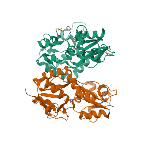
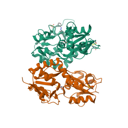
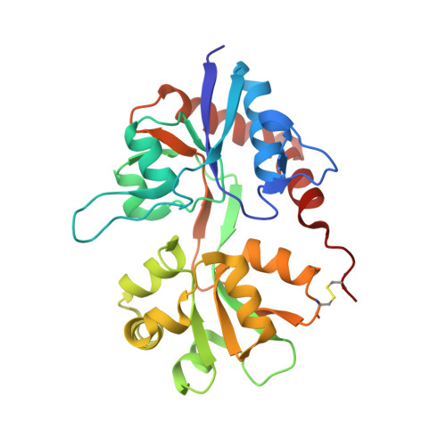
Structure of GluK2-LBD in complex with GluAzo


Experimental Data Snapshot

  • Method: X-RAY DIFFRACTION
  • Resolution: 2.00 Å
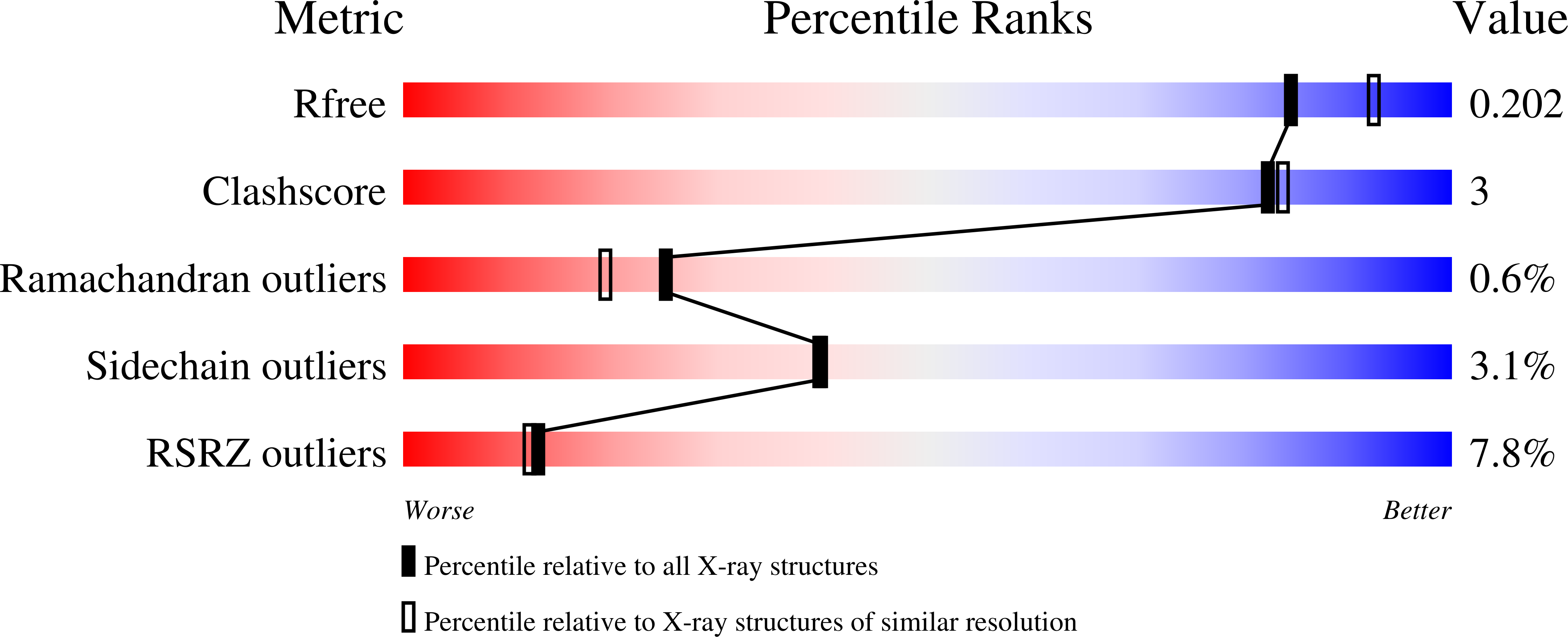
  • R-Value Free: 
    0.204 (Depositor), 0.200 (DCC) 
  • R-Value Work: 
    0.173 (Depositor), 0.170 (DCC) 
  • R-Value Observed: 
    0.175 (Depositor) 

Starting Model: experimental
View more details

wwPDB Validation   3D Report Full Report


Ligand Structure Quality Assessment 

Created with Raphaël 2.3.0Worse 01 BetterLigand structure goodness of fit to experimental dataBest fitted 11WClick on this verticalbar to view details

This is version 1.5 of the entry. See complete history


Literature

A photoswitchable neurotransmitter analogue bound to its receptor.

Reiter, A.Skerra, A.Trauner, D.Schiefner, A.

(2013) Biochemistry 52: 8972-8974

  • DOI: https://doi.org/10.1021/bi4014402
  • Primary Citation of Related Structures:  
    4H8I

  • PubMed Abstract: 

    Incorporation of the azobenzene derivative gluazo, a synthetic photochromic ligand, into a kainate receptor allows for the optical control of neuronal activity. The crystal structure of gluazo bound to a dimeric GluK2 ligand-binding domain reveals one monomer in a closed conformation, occupied by gluazo, and the other in an open conformation, with a bound buffer molecule. The glutamate group of gluazo interacts like the natural glutamate ligand, while its trans-azobenzene moiety protrudes into a tunnel. This elongated cavity presumably cannot accommodate a cis-azobenzene, which explains the reversible activation of the receptor upon photoisomerization.


  • Organizational Affiliation

    Munich Center for Integrated Protein Science (CIPS-M).


Macromolecules
Find similar proteins by:  (by identity cutoff)  |  3D Structure
Entity ID: 1
MoleculeChains Sequence LengthOrganismDetailsImage
Glutamate receptor, ionotropic kainate 2
A, B
259Rattus norvegicusMutation(s): 0 
Gene Names: Glur6Grik2
UniProt
Find proteins for P42260 (Rattus norvegicus)
Explore P42260 
Go to UniProtKB:  P42260
Entity Groups  
Sequence Clusters30% Identity50% Identity70% Identity90% Identity95% Identity100% Identity
UniProt GroupP42260
Sequence Annotations
Expand
  • Reference Sequence
Experimental Data & Validation

Experimental Data

  • Method: X-RAY DIFFRACTION
  • Resolution: 2.00 Å
  • R-Value Free:  0.204 (Depositor), 0.200 (DCC) 
  • R-Value Work:  0.173 (Depositor), 0.170 (DCC) 
  • R-Value Observed: 0.175 (Depositor) 
Space Group: P 61 2 2
Unit Cell:
Length ( Å )Angle ( ˚ )
a = 102.609α = 90
b = 102.609β = 90
c = 282.513γ = 120
Software Package:
Software NamePurpose
XSCALEdata scaling
PHASERphasing
REFMACrefinement
PDB_EXTRACTdata extraction
MAR345data collection
XDSdata reduction

Structure Validation

View Full Validation Report


Ligand Structure Quality Assessment 

Created with Raphaël 2.3.0Worse 01 BetterLigand structure goodness of fit to experimental dataBest fitted 11WClick on this verticalbar to view details

Entry History 

Deposition Data

Revision History  (Full details and data files)

  • Version 1.0: 2013-09-25
    Type: Initial release
  • Version 1.1: 2014-12-24
    Changes: Database references
  • Version 1.2: 2017-07-26
    Changes: Refinement description, Source and taxonomy
  • Version 1.3: 2017-11-15
    Changes: Refinement description
  • Version 1.4: 2023-09-20
    Changes: Data collection, Database references, Derived calculations, Refinement description
  • Version 1.5: 2024-11-27
    Changes: Structure summary