3ZZM | pdb_00003zzm

Crystal structure of Mycobacterium tuberculosis PurH with a novel bound nucleotide CFAIR, at 2.2 A resolution.


Experimental Data Snapshot

  • Method: X-RAY DIFFRACTION
  • Resolution: 2.20 Å
  • R-Value Free: 
    0.235 (Depositor), 0.230 (DCC) 
  • R-Value Work: 
    0.198 (Depositor), 0.190 (DCC) 
  • R-Value Observed: 
    0.200 (Depositor) 

Starting Models: experimental
View more details

wwPDB Validation   3D Report Full Report


Ligand Structure Quality Assessment 

Created with Raphaël 2.3.0Worse 01 BetterLigand structure goodness of fit to experimental dataBest fitted JLNClick on this verticalbar to view details

This is version 1.4 of the entry. See complete history


Literature

Structural Analyses of a Purine Biosynthetic Enzyme from Mycobacterium Tuberculosis Reveal a Novel Bound Nucleotide.

Le Nours, J.Bulloch, E.M.M.Zhang, Z.Greenwood, D.R.Middleditch, M.J.Dickson, J.M.J.Baker, E.N.

(2011) J Biological Chem 286: 40706

  • DOI: https://doi.org/10.1074/jbc.M111.291138
  • Primary Citation of Related Structures:  
    3ZZM, 4A1O

  • PubMed Abstract: 

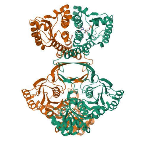
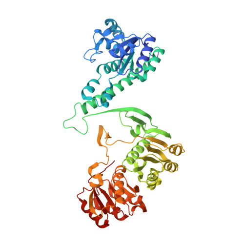
    Enzymes of the de novo purine biosynthetic pathway have been identified as essential for the growth and survival of Mycobacterium tuberculosis and thus have potential for the development of anti-tuberculosis drugs. The final two steps of this pathway are carried out by the bifunctional enzyme 5-aminoimidazole-4-carboxamide ribonucleotide transformylase/inosine monophosphate cyclohydrolase (ATIC), also known as PurH. This enzyme has already been the target of anti-cancer drug development. We have determined the crystal structures of the M. tuberculosis ATIC (Rv0957) both with and without the substrate 5-aminoimidazole-4-carboxamide ribonucleotide, at resolutions of 2.5 and 2.2 Å, respectively. As for other ATIC enzymes, the protein is folded into two domains, the N-terminal domain (residues 1-212) containing the cyclohydrolase active site and the C-terminal domain (residues 222-523) containing the formyltransferase active site. An adventitiously bound nucleotide was found in the cyclohydrolase active site in both structures and was identified by NMR and mass spectral analysis as a novel 5-formyl derivative of an earlier intermediate in the biosynthetic pathway 4-carboxy-5-aminoimidazole ribonucleotide. This result and other studies suggest that this novel nucleotide is a cyclohydrolase inhibitor. The dimer formed by M. tuberculosis ATIC is different from those seen for human and avian ATICs, but it has a similar ∼50-Å separation of the two active sites of the bifunctional enzyme. Evidence in M. tuberculosis ATIC for reactivity of half-the-sites in the cyclohydrolase domains can be attributed to ligand-induced movements that propagate across the dimer interface and may be a common feature of ATIC enzymes.


  • Organizational Affiliation

    Maurice Wilkins Centre for Molecular Biodiscovery and School of Biological Sciences, University of Auckland, Private Bag 92019, Auckland, New Zealand.


Macromolecules
Find similar proteins by:  (by identity cutoff)  |  3D Structure
Entity ID: 1
MoleculeChains Sequence LengthOrganismDetailsImage
BIFUNCTIONAL PURINE BIOSYNTHESIS PROTEIN PURH
A, B
523Mycobacterium tuberculosis H37RvMutation(s): 0 
EC: 2.1.2.3 (PDB Primary Data), 3.5.4.10 (PDB Primary Data)
UniProt
Find proteins for P9WHM7 (Mycobacterium tuberculosis (strain ATCC 25618 / H37Rv))
Explore P9WHM7 
Go to UniProtKB:  P9WHM7
Entity Groups  
Sequence Clusters30% Identity50% Identity70% Identity90% Identity95% Identity100% Identity
UniProt GroupP9WHM7
Sequence Annotations
Expand
  • Reference Sequence
Small Molecules
Ligands 4 Unique
IDChains Name / Formula / InChI Key2D Diagram3D Interactions
JLN
Query on JLN

Download Ideal Coordinates CCD File 
E [auth A]5-(FORMYLAMINO)-1-(5-O-PHOSPHONO-BETA-D-RIBOFURANOSYL)-1H-IMIDAZOLE-4-CARBOXYLIC ACID
C10 H14 N3 O10 P
UEBVPQPUWAADHX-FJGDRVTGSA-N
PO4
Query on PO4

Download Ideal Coordinates CCD File 
C [auth A]PHOSPHATE ION
O4 P
NBIIXXVUZAFLBC-UHFFFAOYSA-K
GOL
Query on GOL

Download Ideal Coordinates CCD File 
G [auth B]GLYCEROL
C3 H8 O3
PEDCQBHIVMGVHV-UHFFFAOYSA-N
K
Query on K

Download Ideal Coordinates CCD File 
D [auth A],
F [auth B]
POTASSIUM ION
K
NPYPAHLBTDXSSS-UHFFFAOYSA-N
Experimental Data & Validation

Experimental Data

  • Method: X-RAY DIFFRACTION
  • Resolution: 2.20 Å
  • R-Value Free:  0.235 (Depositor), 0.230 (DCC) 
  • R-Value Work:  0.198 (Depositor), 0.190 (DCC) 
  • R-Value Observed: 0.200 (Depositor) 
Space Group: P 21 21 21
Unit Cell:
Length ( Å )Angle ( ˚ )
a = 76.594α = 90
b = 108.22β = 90
c = 130.634γ = 90
Software Package:
Software NamePurpose
BUSTERrefinement
HKL-2000data reduction
SCALEPACKdata scaling
PHASERphasing

Structure Validation

View Full Validation Report


Ligand Structure Quality Assessment 

Created with Raphaël 2.3.0Worse 01 BetterLigand structure goodness of fit to experimental dataBest fitted JLNClick on this verticalbar to view details

Entry History 

Deposition Data

Revision History  (Full details and data files)

  • Version 1.0: 2011-09-28
    Type: Initial release
  • Version 1.1: 2011-10-26
    Changes: Database references, Other
  • Version 1.2: 2011-12-14
    Changes: Database references
  • Version 1.3: 2013-11-27
    Changes: Source and taxonomy
  • Version 1.4: 2023-12-20
    Changes: Data collection, Database references, Derived calculations, Other, Refinement description