3ZYC | pdb_00003zyc

DYNAMIN 1 GTPASE GED FUSION DIMER COMPLEXED WITH GMPPCP


Experimental Data Snapshot

  • Method: X-RAY DIFFRACTION
  • Resolution: 2.20 Å
  • R-Value Free: 
    0.269 (Depositor), 0.270 (DCC) 
  • R-Value Work: 
    0.225 (Depositor), 0.220 (DCC) 
  • R-Value Observed: 
    0.225 (Depositor) 

Starting Model: experimental
View more details

wwPDB Validation   3D Report Full Report


Ligand Structure Quality Assessment 

Created with Raphaël 2.3.0Worse 01 BetterLigand structure goodness of fit to experimental dataBest fitted GCPClick on this verticalbar to view details

This is version 1.4 of the entry. See complete history


Literature

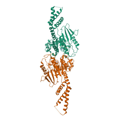
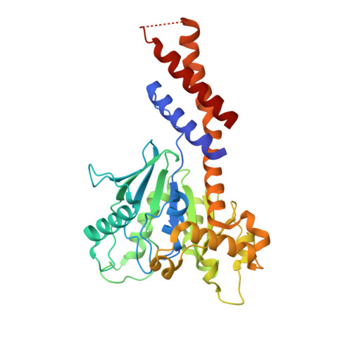
A Pseudoatomic Model of the Dynamin Polymer Identifies a Hydrolysis-Dependent Powerstroke.

Chappie, J.S.Mears, J.A.Fang, S.Leonard, M.Schmid, S.L.Milligan, R.A.Hinshaw, J.E.Dyda, F.

(2011) Cell 147: 209

  • DOI: https://doi.org/10.1016/j.cell.2011.09.003
  • Primary Citation of Related Structures:  
    3ZYC, 3ZYS

  • PubMed Abstract: 

    The GTPase dynamin catalyzes membrane fission by forming a collar around the necks of clathrin-coated pits, but the specific structural interactions and conformational changes that drive this process remain a mystery. We present the GMPPCP-bound structures of the truncated human dynamin 1 helical polymer at 12.2 Å and a fusion protein, GG, linking human dynamin 1's catalytic G domain to its GTPase effector domain (GED) at 2.2 Å. The structures reveal the position and connectivity of dynamin fragments in the assembled structure, showing that G domain dimers only form between tetramers in sequential rungs of the dynamin helix. Using chemical crosslinking, we demonstrate that dynamin tetramers are made of two dimers, in which the G domain of one molecule interacts in trans with the GED of another. Structural comparison of GG(GMPPCP) to the GG transition-state complex identifies a hydrolysis-dependent powerstroke that may play a role in membrane-remodeling events necessary for fission.


  • Organizational Affiliation

    Laboratory of Molecular Biology, National Institute of Diabetes and Digestive and Kidney Diseases, NIH, Bethesda, MD 20892, USA.


Macromolecules
Find similar proteins by:  (by identity cutoff)  |  3D Structure
Entity ID: 1
MoleculeChains Sequence LengthOrganismDetailsImage
DYNAMIN-1A,
B [auth D]
353Homo sapiensMutation(s): 1 
EC: 3.6.5.5
UniProt & NIH Common Fund Data Resources
Find proteins for Q05193 (Homo sapiens)
Explore Q05193 
Go to UniProtKB:  Q05193
PHAROS:  Q05193
GTEx:  ENSG00000106976 
Entity Groups  
Sequence Clusters30% Identity50% Identity70% Identity90% Identity95% Identity100% Identity
UniProt GroupQ05193
Sequence Annotations
Expand
  • Reference Sequence
Experimental Data & Validation

Experimental Data

  • Method: X-RAY DIFFRACTION
  • Resolution: 2.20 Å
  • R-Value Free:  0.269 (Depositor), 0.270 (DCC) 
  • R-Value Work:  0.225 (Depositor), 0.220 (DCC) 
  • R-Value Observed: 0.225 (Depositor) 
Space Group: P 1 21 1
Unit Cell:
Length ( Å )Angle ( ˚ )
a = 44.151α = 90
b = 82.635β = 96.68
c = 95.014γ = 90
Software Package:
Software NamePurpose
CNSrefinement
XDSdata reduction
XDSdata scaling
PHASERphasing

Structure Validation

View Full Validation Report


Ligand Structure Quality Assessment 

Created with Raphaël 2.3.0Worse 01 BetterLigand structure goodness of fit to experimental dataBest fitted GCPClick on this verticalbar to view details

Entry History 

Deposition Data

Revision History  (Full details and data files)

  • Version 1.0: 2011-10-12
    Type: Initial release
  • Version 1.1: 2017-06-28
    Changes: Data collection
  • Version 1.2: 2019-05-08
    Changes: Data collection, Experimental preparation
  • Version 1.3: 2019-05-15
    Changes: Data collection, Experimental preparation
  • Version 1.4: 2023-12-20
    Changes: Data collection, Database references, Derived calculations, Other, Refinement description