3JZT | pdb_00003jzt

Structure of a cubic crystal form of X (ADRP) domain from FCoV with ADP-ribose


Experimental Data Snapshot

  • Method: X-RAY DIFFRACTION
  • Resolution: 3.91 Å
  • R-Value Free: 
    0.239 (Depositor), 0.240 (DCC) 
  • R-Value Work: 
    0.199 (Depositor), 0.200 (DCC) 
  • R-Value Observed: 
    0.199 (Depositor) 

wwPDB Validation   3D Report Full Report


Ligand Structure Quality Assessment 

Created with Raphaël 2.3.0Worse 01 BetterLigand structure goodness of fit to experimental dataBest fitted APRClick on this verticalbar to view details

This is version 1.2 of the entry. See complete history


Literature

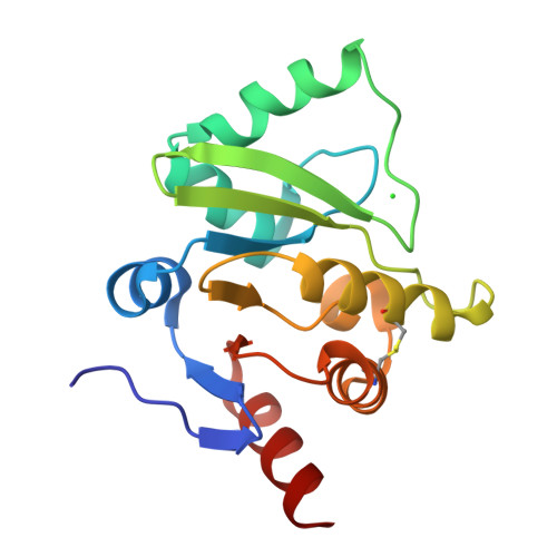
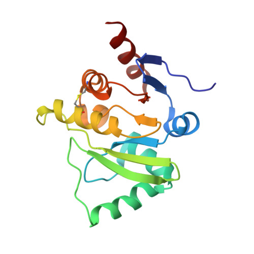
Structure of the X (ADRP) domain of nsp3 from feline coronavirus

Wojdyla, J.A.Manolaridis, I.Snijder, E.J.Gorbalenya, A.E.Coutard, B.Piotrowski, Y.Hilgenfeld, R.Tucker, P.A.

(2009) Acta Crystallogr D Biol Crystallogr 65: 1292-1300

  • DOI: https://doi.org/10.1107/S0907444909040074
  • Primary Citation of Related Structures:  
    3ETI, 3EW5, 3JZT

  • PubMed Abstract: 

    The structure of the X (or ADRP) domain of a pathogenic variant of feline coronavirus (FCoV) has been determined in tetragonal and cubic crystal forms to 3.1 and 2.2 A resolution, respectively. In the tetragonal crystal form, glycerol-3-phosphate was observed in the ADP-ribose-binding site. Both crystal forms contained large solvent channels and had a solvent content of higher than 70%. Only very weak binding of this domain to ADP-ribose was detected in vitro. However, the structure with ADP-ribose bound was determined in the cubic crystal form at 3.9 A resolution. The structure of the FCoV X domain had the expected macro-domain fold and is the first structure of this domain from a coronavirus belonging to subgroup 1a.


  • Organizational Affiliation

    EMBL Hamburg Outstation, c/o DESY, D-22603 Hamburg, Germany.


Macromolecules
Find similar proteins by:  (by identity cutoff)  |  3D Structure
Entity ID: 1
MoleculeChains Sequence LengthOrganismDetailsImage
macro domain of Non-structural protein 3
A, B, C, D, E
168Feline infectious peritonitis virus (strain 79-1146)Mutation(s): 0 
Gene Names: rep1a-1b
UniProt
Find proteins for Q98VG9 (Feline coronavirus (strain FIPV WSU-79/1146))
Explore Q98VG9 
Go to UniProtKB:  Q98VG9
Entity Groups  
Sequence Clusters30% Identity50% Identity70% Identity90% Identity95% Identity100% Identity
UniProt GroupQ98VG9
Sequence Annotations
Expand
  • Reference Sequence
Experimental Data & Validation

Experimental Data

  • Method: X-RAY DIFFRACTION
  • Resolution: 3.91 Å
  • R-Value Free:  0.239 (Depositor), 0.240 (DCC) 
  • R-Value Work:  0.199 (Depositor), 0.200 (DCC) 
  • R-Value Observed: 0.199 (Depositor) 
Space Group: P 21 3
Unit Cell:
Length ( Å )Angle ( ˚ )
a = 220.236α = 90
b = 220.236β = 90
c = 220.236γ = 90
Software Package:
Software NamePurpose
SCALAdata scaling
REFMACrefinement
PDB_EXTRACTdata extraction
XDSdata scaling
COMBATdata reduction
Auto-Rickshawphasing

Structure Validation

View Full Validation Report


Ligand Structure Quality Assessment 

Created with Raphaël 2.3.0Worse 01 BetterLigand structure goodness of fit to experimental dataBest fitted APRClick on this verticalbar to view details

Entry History 

Deposition Data

Revision History  (Full details and data files)

  • Version 1.0: 2010-01-12
    Type: Initial release
  • Version 1.1: 2011-07-13
    Changes: Version format compliance
  • Version 1.2: 2024-11-20
    Changes: Data collection, Database references, Derived calculations, Refinement description, Structure summary