2G9X | pdb_00002g9x

Structure of Thr 160 phosphorylated CDK2/cyclin A in complex with the inhibitor NU6271


Experimental Data Snapshot

  • Method: X-RAY DIFFRACTION
  • Resolution: 2.50 Å
  • R-Value Free: 
    0.265 (Depositor), 0.260 (DCC) 
  • R-Value Work: 
    0.217 (Depositor), 0.220 (DCC) 
  • R-Value Observed: 
    0.219 (Depositor) 

Starting Model: experimental
View more details

wwPDB Validation   3D Report Full Report


Ligand Structure Quality Assessment 

Created with Raphaël 2.3.0Worse 01 BetterLigand structure goodness of fit to experimental dataBest fitted NU5Click on this verticalbar to view details

This is version 1.4 of the entry. See complete history


Literature

Searching for Cyclin-Dependent Kinase Inhibitors Using a New Variant of the Cope Elimination.

Griffin, R.J.Henderson, A.Curtin, N.J.Echalier, A.Endicott, J.A.Hardcastle, I.R.Newell, D.R.Noble, M.E.Wang, L.Z.Golding, B.T.

(2006) J Am Chem Soc 128: 6012-6013

  • DOI: https://doi.org/10.1021/ja060595j
  • Primary Citation of Related Structures:  
    2G9X

  • PubMed Abstract: 

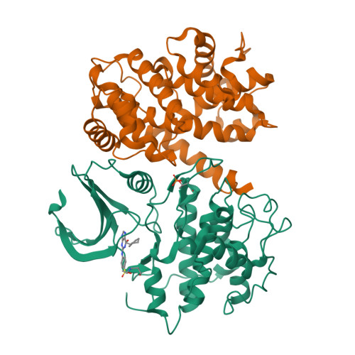
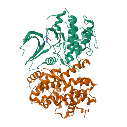
    beta-Piperidinoethylsulfides are oxidized by m-chloroperbenzoic acid to intermediates containing both N-oxide and sulfone functions. These undergo a Cope-type elimination to a vinylsulfone that can be captured by amines to afford beta-aminoethylsulfones. When a beta-aminoethylsulfone group is linked to the 4-position of a phenyl group attached at N-2 of O6-cyclohexylmethylguanine, the resulting derivatives are inhibitors of the cyclin-dependent kinase CDK2. One of the most potent inhibitors (IC50 = 45 nM) contained a N-3-hydroxypropyl group on the aminoethylsulfonyl substituent. The crystal structure of this inhibitor bound to CDK2/cyclin A was determined and shows an unusual network of hydrogen bonds. The synthetic methodology developed can be utilized in multiple-parallel format and has numerous potential applications in medicinal chemistry.


  • Organizational Affiliation

    Northern Institute for Cancer Research, School of Natural Sciences-Chemistry, Bedson Building, University of Newcastle, Newcastle upon Tyne, UK.


Macromolecules
Find similar proteins by:  (by identity cutoff)  |  3D Structure
Entity ID: 1
MoleculeChains Sequence LengthOrganismDetailsImage
Cell division protein kinase 2
A, C
299Homo sapiensMutation(s): 1 
Gene Names: CDK2
EC: 2.7.1.37 (PDB Primary Data), 2.7.11.22 (UniProt)
UniProt & NIH Common Fund Data Resources
Find proteins for P24941 (Homo sapiens)
Explore P24941 
Go to UniProtKB:  P24941
PHAROS:  P24941
GTEx:  ENSG00000123374 
Entity Groups  
Sequence Clusters30% Identity50% Identity70% Identity90% Identity95% Identity100% Identity
UniProt GroupP24941
Sequence Annotations
Expand
  • Reference Sequence
Find similar proteins by:  (by identity cutoff)  |  3D Structure
Entity ID: 2
MoleculeChains Sequence LengthOrganismDetailsImage
Cyclin-A2
B, D
262Bos taurusMutation(s): 0 
Gene Names: CCNA2CCNA
UniProt
Find proteins for P30274 (Bos taurus)
Explore P30274 
Go to UniProtKB:  P30274
Entity Groups  
Sequence Clusters30% Identity50% Identity70% Identity90% Identity95% Identity100% Identity
UniProt GroupP30274
Sequence Annotations
Expand
  • Reference Sequence
Small Molecules
Ligands 1 Unique
IDChains Name / Formula / InChI Key2D Diagram3D Interactions
NU5
Query on NU5

Download Ideal Coordinates CCD File 
E [auth A],
F [auth C]
3-({2-[(4-{[6-(CYCLOHEXYLMETHOXY)-9H-PURIN-2-YL]AMINO}PHENYL)SULFONYL]ETHYL}AMINO)PROPAN-1-OL
C23 H32 N6 O4 S
AMFGILNPFBVREA-UHFFFAOYSA-N
Modified Residues  1 Unique
IDChains TypeFormula2D DiagramParent
TPO
Query on TPO
A, C
L-PEPTIDE LINKINGC4 H10 N O6 PTHR
Experimental Data & Validation

Experimental Data

  • Method: X-RAY DIFFRACTION
  • Resolution: 2.50 Å
  • R-Value Free:  0.265 (Depositor), 0.260 (DCC) 
  • R-Value Work:  0.217 (Depositor), 0.220 (DCC) 
  • R-Value Observed: 0.219 (Depositor) 
Space Group: P 21 21 21
Unit Cell:
Length ( Å )Angle ( ˚ )
a = 74.58α = 90
b = 134.986β = 90
c = 148.513γ = 90
Software Package:
Software NamePurpose
REFMACrefinement
MOSFLMdata reduction
CCP4data scaling
MOLREPphasing

Structure Validation

View Full Validation Report


Ligand Structure Quality Assessment 

Created with Raphaël 2.3.0Worse 01 BetterLigand structure goodness of fit to experimental dataBest fitted NU5Click on this verticalbar to view details

Entry History 

Deposition Data

Revision History  (Full details and data files)

  • Version 1.0: 2006-05-23
    Type: Initial release
  • Version 1.1: 2008-05-01
    Changes: Version format compliance
  • Version 1.2: 2011-07-13
    Changes: Advisory, Version format compliance
  • Version 1.3: 2023-08-30
    Changes: Data collection, Database references, Derived calculations, Refinement description
  • Version 1.4: 2024-11-20
    Changes: Structure summary