1TQ2 | pdb_00001tq2

Crystal Structure of IIGP1: a paradigm for interferon inducible p47 resistance GTPases


Experimental Data Snapshot

  • Method: X-RAY DIFFRACTION
  • Resolution: 2.70 Å
  • R-Value Free: 
    0.280 (Depositor), 0.270 (DCC) 
  • R-Value Work: 
    0.222 (Depositor), 0.210 (DCC) 
  • R-Value Observed: 
    0.240 (Depositor) 

wwPDB Validation   3D Report Full Report


Ligand Structure Quality Assessment 

Created with Raphaël 2.3.0Worse 01 BetterLigand structure goodness of fit to experimental dataBest fitted GNPClick on this verticalbar to view details

This is version 1.3 of the entry. See complete history


Literature

Crystal Structure of IIGP1; A Paradigm for Interferon-Inducible p47 Resistance GTPases

Ghosh, A.Uthaiah, R.Howard, J.Herrmann, C.Wolf, E.

(2004) Mol Cell 15: 727-739

  • DOI: https://doi.org/10.1016/j.molcel.2004.07.017
  • Primary Citation of Related Structures:  
    1TPZ, 1TQ2, 1TQ4, 1TQ6, 1TQD

  • PubMed Abstract: 

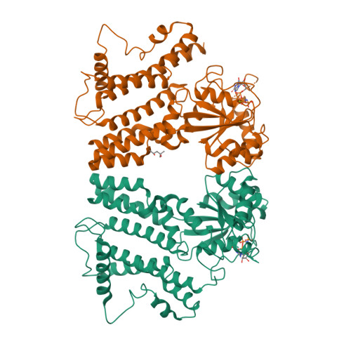
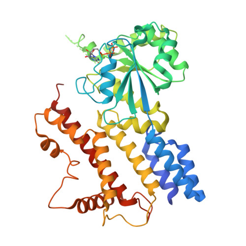
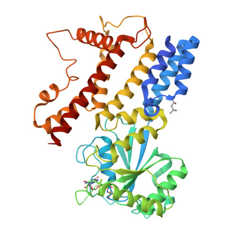
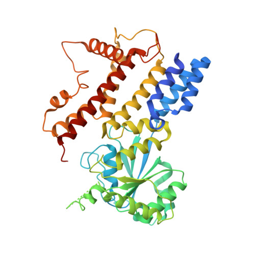
    Interferon-inducible p47 GTPases are critical mediators of cell-autonomous resistance against several intracellular pathogens. Here we present the first crystal structure of a member of this novel GTPase family, IIGP1, in its nucleotide-free, GDP-, and GppNHp-bound form. The structure shows a Ras-like G domain between an N-terminal three-helix bundle and a complex system of C-terminal helices and loops. Sequence comparison and secondary structure prediction suggest the IIGP1 structure to be a valid model for the p47 GTPase family. The IIGP1 crystals contain a noncrystallographic dimer. We show that the dimer is required for cooperative GTP hydrolysis and GTP-dependent oligomerization of IIGP1. We also present the GDP- and GppNHp-bound monomeric structures of two dimer interface mutants. Our structures direct approaches to the analysis of the catalytic mechanism of IIGP1 and provide a coherent basis for structure-function studies aimed at elucidating the mechanistic basis of pathogen resistance caused by these enigmatic GTPases.


  • Organizational Affiliation

    Department of Structural Biology, Max Planck Institute for Molecular Physiology, Otto-Hahn-Strasse 11, 44227 Dortmund, Germany.


Macromolecules
Find similar proteins by:  (by identity cutoff)  |  3D Structure
Entity ID: 1
MoleculeChains Sequence LengthOrganismDetailsImage
interferon-inducible GTPase
A, B
422Mus musculusMutation(s): 0 
Gene Names: IIGP1
EC: 3.6.5
UniProt
Find proteins for Q9QZ85 (Mus musculus)
Explore Q9QZ85 
Go to UniProtKB:  Q9QZ85
Entity Groups  
Sequence Clusters30% Identity50% Identity70% Identity90% Identity95% Identity100% Identity
UniProt GroupQ9QZ85
Sequence Annotations
Expand
  • Reference Sequence
Experimental Data & Validation

Experimental Data

  • Method: X-RAY DIFFRACTION
  • Resolution: 2.70 Å
  • R-Value Free:  0.280 (Depositor), 0.270 (DCC) 
  • R-Value Work:  0.222 (Depositor), 0.210 (DCC) 
  • R-Value Observed: 0.240 (Depositor) 
Space Group: P 21 21 21
Unit Cell:
Length ( Å )Angle ( ˚ )
a = 81.86α = 90
b = 92.67β = 90
c = 142.4γ = 90
Software Package:
Software NamePurpose
XDSdata scaling
XDSdata reduction
AMoREphasing
CNSrefinement

Structure Validation

View Full Validation Report


Ligand Structure Quality Assessment 

Created with Raphaël 2.3.0Worse 01 BetterLigand structure goodness of fit to experimental dataBest fitted GNPClick on this verticalbar to view details

Entry History 

Deposition Data

Revision History  (Full details and data files)

  • Version 1.0: 2004-09-21
    Type: Initial release
  • Version 1.1: 2008-04-30
    Changes: Version format compliance
  • Version 1.2: 2011-07-13
    Changes: Version format compliance
  • Version 1.3: 2024-03-13
    Changes: Data collection, Database references, Derived calculations