1JVP | pdb_00001jvp

Crystal structure of human CDK2 (unphosphorylated) in complex with PKF049-365


Experimental Data Snapshot

  • Method: X-RAY DIFFRACTION
  • Resolution: 1.53 Å
  • R-Value Free: 
    0.253 (Depositor), 0.240 (DCC) 
  • R-Value Work: 
    0.206 (Depositor), 0.200 (DCC) 
  • R-Value Observed: 
    0.210 (Depositor) 

wwPDB Validation   3D Report Full Report


Ligand Structure Quality Assessment 

Created with Raphaël 2.3.0Worse 01 BetterLigand structure goodness of fit to experimental dataBest fitted 89EClick on this verticalbar to view details

This is version 2.1 of the entry. See complete history


Literature

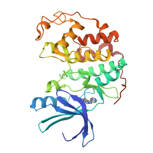
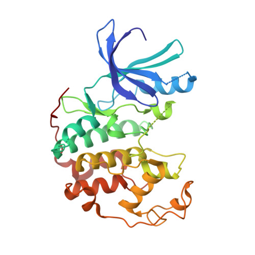
Structure-based design and protein X-ray analysis of a protein kinase inhibitor.

Furet, P.Meyer, T.Strauss, A.Raccuglia, S.Rondeau, J.M.

(2002) Bioorg Med Chem Lett 12: 221-224

  • DOI: https://doi.org/10.1016/s0960-894x(01)00715-6
  • Primary Citation of Related Structures:  
    1JVP

  • PubMed Abstract: 

    A 5-aryl-1H-pyrazole molecular scaffold was designed to ligate the ATP binding site of cyclin dependent kinase 2 (CDK2) on the basis of crystallographic information. A search of the compound collection of Novartis using this scaffold as substructure query led to the identification of PKF049-365 as a representative of a new class of inhibitors of the cell cycle kinases CDK1/2. The three-dimensional structure of CDK2 in complex with PKF049-365 was subsequently determined by protein crystallography and refined to 1.53 A resolution. The X-ray analysis confirmed the binding mode expected from the design hypothesis. In addition, it revealed an alternative binding orientation involving a second tautomeric form of the inhibitor that was not envisaged during the design stage.


  • Organizational Affiliation

    Novartis Pharmaceuticals Inc., Oncology Research, CH-4002 Basel, Switzerland. pascal.furet@pharma.novartis.com


Macromolecules
Find similar proteins by:  (by identity cutoff)  |  3D Structure
Entity ID: 1
MoleculeChains Sequence LengthOrganismDetailsImage
Cell division protein kinase 2A [auth P]298Homo sapiensMutation(s): 0 
EC: 2.7.1.37 (PDB Primary Data), 2.7.11.22 (UniProt)
UniProt & NIH Common Fund Data Resources
Find proteins for P24941 (Homo sapiens)
Explore P24941 
Go to UniProtKB:  P24941
PHAROS:  P24941
GTEx:  ENSG00000123374 
Entity Groups  
Sequence Clusters30% Identity50% Identity70% Identity90% Identity95% Identity100% Identity
UniProt GroupP24941
Sequence Annotations
Expand
  • Reference Sequence
Small Molecules
Ligands 1 Unique
IDChains Name / Formula / InChI Key2D Diagram3D Interactions
89E
Query on 89E

Download Ideal Coordinates CCD File 
B [auth P]3-pyridin-4-yl-2,4-dihydroindeno[1,2-c]pyrazole
C15 H11 N3
NHOACLCXCKJMAK-UHFFFAOYSA-N
Experimental Data & Validation

Experimental Data

  • Method: X-RAY DIFFRACTION
  • Resolution: 1.53 Å
  • R-Value Free:  0.253 (Depositor), 0.240 (DCC) 
  • R-Value Work:  0.206 (Depositor), 0.200 (DCC) 
  • R-Value Observed: 0.210 (Depositor) 
Space Group: P 21 21 21
Unit Cell:
Length ( Å )Angle ( ˚ )
a = 53.528α = 90
b = 71.802β = 90
c = 72.362γ = 90
Software Package:
Software NamePurpose
AMoREphasing
X-PLORrefinement
DENZOdata reduction
SCALEPACKdata scaling

Structure Validation

View Full Validation Report


Ligand Structure Quality Assessment 

Created with Raphaël 2.3.0Worse 01 BetterLigand structure goodness of fit to experimental dataBest fitted 89EClick on this verticalbar to view details

Entry History 

Deposition Data

Revision History  (Full details and data files)

  • Version 1.0: 2001-12-21
    Type: Initial release
  • Version 1.1: 2008-04-27
    Changes: Version format compliance
  • Version 1.2: 2011-07-13
    Changes: Version format compliance
  • Version 2.0: 2021-09-22
    Changes: Atomic model, Data collection, Database references, Derived calculations, Non-polymer description, Structure summary
  • Version 2.1: 2024-02-07
    Changes: Data collection