1IRA | pdb_00001ira

COMPLEX OF THE INTERLEUKIN-1 RECEPTOR WITH THE INTERLEUKIN-1 RECEPTOR ANTAGONIST (IL1RA)


Experimental Data Snapshot

  • Method: X-RAY DIFFRACTION
  • Resolution: 2.70 Å
  • R-Value Free: 
    0.314 (Depositor) 
  • R-Value Work: 
    0.213 (Depositor), 0.200 (DCC) 
  • R-Value Observed: 
    0.213 (Depositor) 

Starting Models: experimental
View more details

wwPDB Validation   3D Report Full Report


Ligand Structure Quality Assessment 

Created with Raphaël 2.3.0Worse 01 BetterLigand structure goodness of fit to experimental dataBest fitted NAGClick on this verticalbar to view details

This is version 1.6 of the entry. See complete history


Literature

A new cytokine-receptor binding mode revealed by the crystal structure of the IL-1 receptor with an antagonist.

Schreuder, H.Tardif, C.Trump-Kallmeyer, S.Soffientini, A.Sarubbi, E.Akeson, A.Bowlin, T.Yanofsky, S.Barrett, R.W.

(1997) Nature 386: 194-200

  • DOI: https://doi.org/10.1038/386194a0
  • Primary Citation of Related Structures:  
    1IRA

  • PubMed Abstract: 

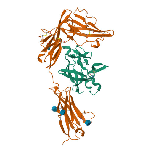
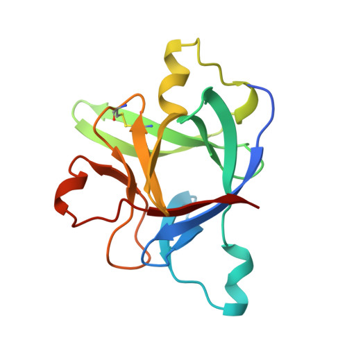
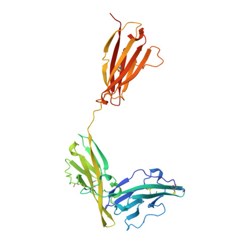
    Inflammation, regardless of whether it is provoked by infection or by tissue damage, starts with the activation of macrophages which initiate a cascade of inflammatory responses by producing the cytokines interleukin-1 (IL-1) and tumour necrosis factor-alpha (ref. 1). Three naturally occurring ligands for the IL-1 receptor (IL1R) exist: the agonists IL-1alpha and IL-1beta and the IL-1-receptor antagonist IL1RA (ref. 2). IL-1 is the only cytokine for which a naturally occurring antagonist is known. Here we describe the crystal structure at 2.7 A resolution of the soluble extracellular part of type-I IL1R complexed with IL1RA. The receptor consists of three immunoglobulin-like domains. Domains 1 and 2 are tightly linked, but domain three is completely separate and connected by a flexible linker. Residues of all three domains contact the antagonist and include the five critical IL1RA residues which were identified by site-directed mutagenesis. A region that is important for biological function in IL-1beta, the 'receptor trigger site' is not in direct contact with the receptor in the IL1RA complex. Modelling studies suggest that this IL-1beta trigger site might induce a movement of domain 3.


  • Organizational Affiliation

    Marion Merrell Dow Research Institute, Strasbourg, France.


Macromolecules
Find similar proteins by:  (by identity cutoff)  |  3D Structure
Entity ID: 1
MoleculeChains Sequence LengthOrganismDetailsImage
INTERLEUKIN-1 RECEPTOR ANTAGONISTA [auth X]152Homo sapiensMutation(s): 0 
UniProt & NIH Common Fund Data Resources
Find proteins for P18510 (Homo sapiens)
Explore P18510 
Go to UniProtKB:  P18510
PHAROS:  P18510
GTEx:  ENSG00000136689 
Entity Groups  
Sequence Clusters30% Identity50% Identity70% Identity90% Identity95% Identity100% Identity
UniProt GroupP18510
Sequence Annotations
Expand
  • Reference Sequence
Find similar proteins by:  (by identity cutoff)  |  3D Structure
Entity ID: 2
MoleculeChains Sequence LengthOrganismDetailsImage
INTERLEUKIN-1 RECEPTORB [auth Y]319Homo sapiensMutation(s): 0 
EC: 3.2.2.6
UniProt & NIH Common Fund Data Resources
Find proteins for P14778 (Homo sapiens)
Explore P14778 
Go to UniProtKB:  P14778
PHAROS:  P14778
GTEx:  ENSG00000115594 
Entity Groups  
Sequence Clusters30% Identity50% Identity70% Identity90% Identity95% Identity100% Identity
UniProt GroupP14778
Glycosylation
Glycosylation Sites: 4Go to GlyGen: P14778-1
Sequence Annotations
Expand
  • Reference Sequence
Experimental Data & Validation

Experimental Data

  • Method: X-RAY DIFFRACTION
  • Resolution: 2.70 Å
  • R-Value Free:  0.314 (Depositor) 
  • R-Value Work:  0.213 (Depositor), 0.200 (DCC) 
  • R-Value Observed: 0.213 (Depositor) 
Space Group: P 21 21 21
Unit Cell:
Length ( Å )Angle ( ˚ )
a = 47.2α = 90
b = 84.6β = 90
c = 140.2γ = 90
Software Package:
Software NamePurpose
XDSdata scaling
XSCALEdata scaling
SQUASHphasing
X-PLORmodel building
X-PLORrefinement
XDSdata reduction
X-PLOR3.1phasing

Structure Validation

View Full Validation Report


Ligand Structure Quality Assessment 

Created with Raphaël 2.3.0Worse 01 BetterLigand structure goodness of fit to experimental dataBest fitted NAGClick on this verticalbar to view details

Entry History 

Deposition Data

Revision History  (Full details and data files)

  • Version 1.0: 1998-06-17
    Type: Initial release
  • Version 1.1: 2008-03-24
    Changes: Version format compliance
  • Version 1.2: 2011-07-13
    Changes: Non-polymer description, Version format compliance
  • Version 1.3: 2018-04-04
    Changes: Data collection
  • Version 1.4: 2020-07-29
    Type: Remediation
    Reason: Carbohydrate remediation
    Changes: Data collection, Derived calculations, Structure summary
  • Version 1.5: 2023-08-09
    Changes: Database references, Refinement description, Structure summary
  • Version 1.6: 2024-11-20
    Changes: Data collection, Structure summary