9ZZK | pdb_00009zzk

One Lmod2 and incoming actin at the pointed end of F-actin


Experimental Data Snapshot

  • Method: ELECTRON MICROSCOPY
  • Resolution: 4.06 Å
  • Resolution: 4.06 Å
  • Resolution: 4.06 Å
  • Resolution: 4.06 Å
  • Resolution: 4.06 Å
  • Resolution: 4.06 Å
  • Resolution: 4.06 Å
  • Resolution: 4.06 Å
  • Aggregation State: FILAMENT 
  • Reconstruction Method: SINGLE PARTICLE 

Starting Model: experimental
View more details

wwPDB Validation 3D Report Full Report

Validation slider image for 9ZZK

This is version 1.1 of the entry. See complete history

Literature

Leiomodin 2 is a processive pointed-end elongator of actin filaments.

Biswas, S.Larrinaga, T.M.Choubey, S.Gregorio, C.C.Shekhar, S.

(2026) Nat Commun 17

  • DOI: https://doi.org/10.1038/s41467-026-74809-z
  • Primary Citation Related Structures: 
    9ZZK

  • PubMed Abstract: 

    The actin cytoskeleton drives essential processes like cell migration and muscle contraction. While barbed-end polymerization is well-established, pointed-end elongation was long considered impossible in vivo. Here, we demonstrate that Leiomodin 2 (Lmod2), which localizes to thin-filament pointed ends in striated muscle cells, functions as an actin polymerase for pointed-end elongation. Single-molecule and single-filament imaging reveal that Lmod2 remains processively bound to pointed ends in vitro, enabling elongation even in the presence of high profilin concentrations found in the cytoplasm that otherwise would cause depolymerization of free pointed ends. Kinetic analysis indicates that Lmod2-mediated elongation proceeds through a linked two-step mechanism, in which monomer addition is followed by a first-order transition at the Lmod2-bound pointed end that limits elongation at high actin concentrations. Lmod2's activity also persists in the presence of tropomyosin, underscoring its physiological relevance. Both processivity and elongation rate of Lmod2 are dependent on its WH2 domain. Remarkably, human dilated cardiomyopathy-associated mutations in Lmod2 greatly reduce Lmod2's pointed-end elongation activity, providing a potential mechanism for disease progression and supporting a role for Lmod2-mediated polymerization in the formation and maintenance of muscle sarcomeres.


  • Organizational Affiliation
    • Department of Physics, Emory University, Atlanta, GA, USA.

Macromolecule Content 

  • Total Structure Weight: 316.91 kDa 
  • Atom Count: 18,791 
  • Modeled Residue Count: 2,378 
  • Deposited Residue Count: 2,806 
  • Unique protein chains: 2

Macromolecules

Find similar proteins by:|  3D Structure
Entity ID: 1
MoleculeChains  Sequence LengthOrganismDetailsImage
Actin, alpha skeletal muscle
A,
B,
C,
D,
E,
G [auth O]
375Oryctolagus cuniculusMutation(s): 0 
EC: 3.6.4
UniProt
Find proteins for P68135 (Oryctolagus cuniculus)
Explore P68135 
Go to UniProtKB:  P68135
Entity Groups
Sequence Clusters30% Identity50% Identity70% Identity90% Identity95% Identity100% Identity
UniProt GroupP68135
Sequence Annotations
Expand
Reference Sequence
Find similar proteins by:|  3D Structure
Entity ID: 2
MoleculeChains  Sequence LengthOrganismDetailsImage
Leiomodin-2F [auth M]556Homo sapiensMutation(s): 0 
Gene Names: LMOD2
UniProt & NIH Common Fund Data Resources
Find proteins for Q6P5Q4 (Homo sapiens)
Explore Q6P5Q4 
Go to UniProtKB:  Q6P5Q4
PHAROS:  Q6P5Q4
GTEx:  ENSG00000170807 
Entity Groups
Sequence Clusters30% Identity50% Identity70% Identity90% Identity95% Identity100% Identity
UniProt GroupQ6P5Q4
Sequence Annotations
Expand
Reference Sequence

Small Molecules

Ligands 3 Unique
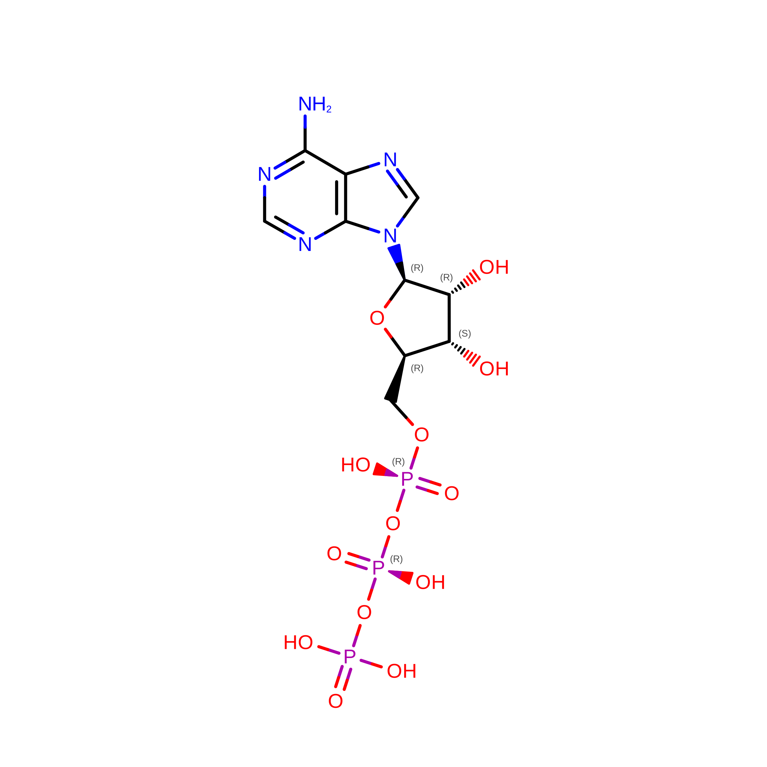
IDChains Name / Formula / InChI Key2D Diagram3D Interactions
ATP

Query on ATP


Download:Ideal Coordinates CCD File
S [auth O]ADENOSINE-5'-TRIPHOSPHATE
C10 H16 N5 O13 P3
ZKHQWZAMYRWXGA-KQYNXXCUSA-N
ADP

Query on ADP


Download:Ideal Coordinates CCD File
H [auth A],
J [auth B],
L [auth C],
N [auth D],
P [auth E]
ADENOSINE-5'-DIPHOSPHATE
C10 H15 N5 O10 P2
XTWYTFMLZFPYCI-KQYNXXCUSA-N
MG

Query on MG


Download:Ideal Coordinates CCD File
I [auth A]
K [auth B]
M [auth C]
O [auth D]
Q [auth E]
I [auth A],
K [auth B],
M [auth C],
O [auth D],
Q [auth E],
R [auth O]
MAGNESIUM ION
Mg
JLVVSXFLKOJNIY-UHFFFAOYSA-N
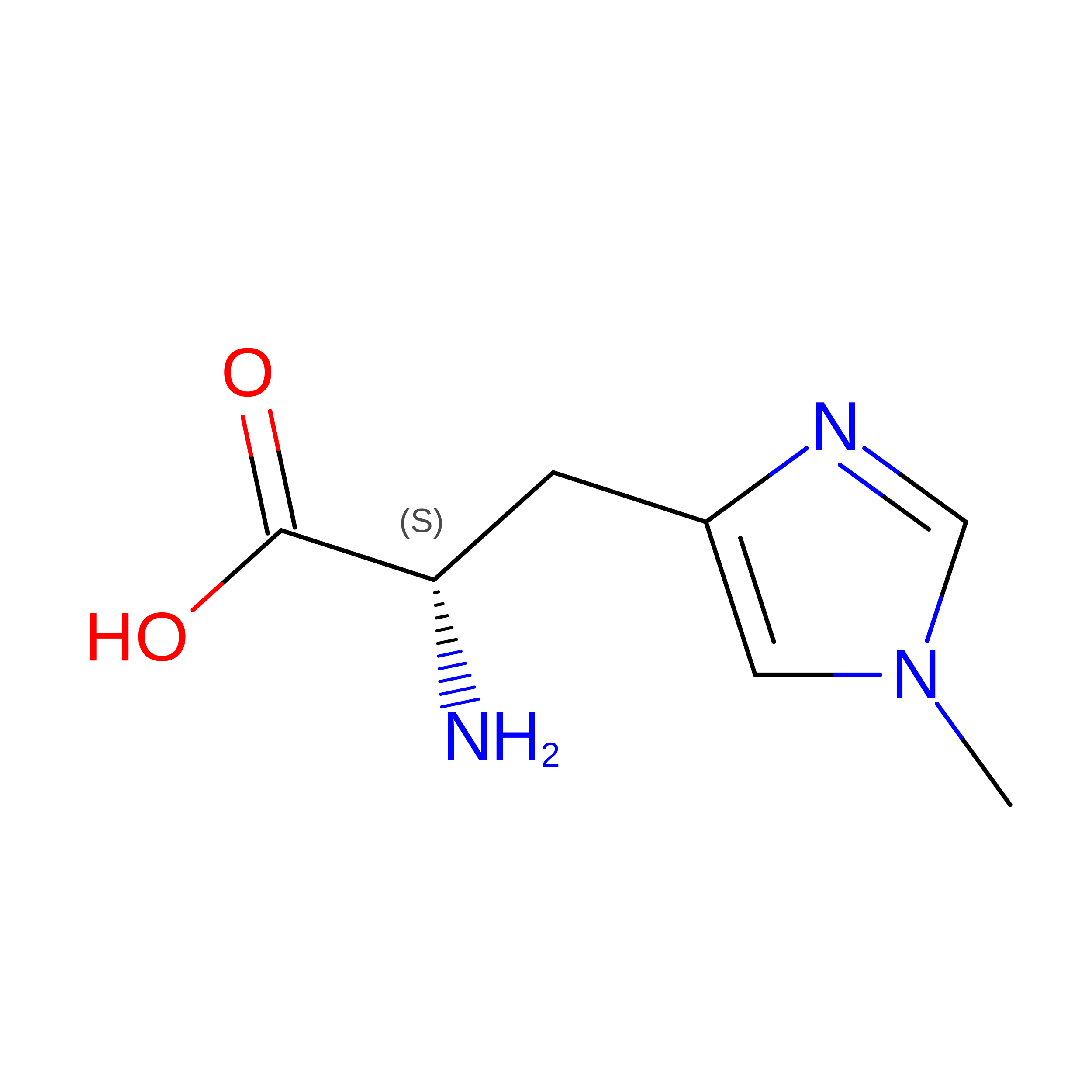
Modified Residues  1 Unique
IDChains TypeFormula2D DiagramParent
HIC
Query on HIC
A
B
C
D
E
A,
B,
C,
D,
E,
G [auth O]
L-PEPTIDE LINKINGC7 H11 N3 O2HIS

Experimental Data & Validation

Experimental Data

  • Method: ELECTRON MICROSCOPY
  • Resolution: 4.06 Å
  • Resolution: 4.06 Å
  • Resolution: 4.06 Å
  • Resolution: 4.06 Å
  • Resolution: 4.06 Å
  • Resolution: 4.06 Å
  • Resolution: 4.06 Å
  • Resolution: 4.06 Å
  • Aggregation State: FILAMENT 
  • Reconstruction Method: SINGLE PARTICLE 
EM Software:
TaskSoftware PackageVersion
RECONSTRUCTIONPHENIX
MODEL REFINEMENTPHENIX1.21.2_5419

Structure Validation

View Full Validation Report


Entry History 

& Funding Information

Deposition Data


Funding OrganizationLocationGrant Number
National Institutes of Health/National Institute of General Medical Sciences (NIH/NIGMS)United States5R01GM152412
National Institutes of Health/National Institute of Arthritis and Musculoskeletal and Skin Diseases (NIH/NIAMS)United States5R01GM073791
National Institutes of Health/National Institute of Arthritis and Musculoskeletal and Skin Diseases (NIH/NIAMS)United States5T32AR053461-20

Revision History  (Full details and data files)

  • Version 1.0: 2026-06-24
    Type: Initial release
  • Version 1.1: 2026-08-05
    Changes: Data collection, Database references