9ZXZ | pdb_00009zxz

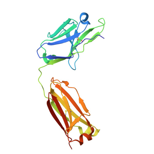
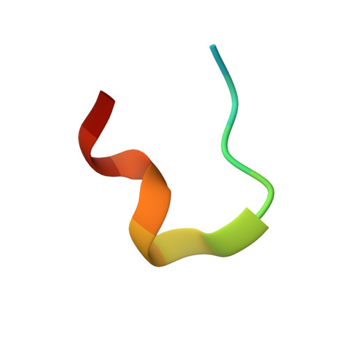
Anti-HCV Fab RM1-38 in complex with E2 HVR1 peptide


Experimental Data Snapshot

  • Method: X-RAY DIFFRACTION
  • Resolution: 2.83 Å
  • R-Value Free: 
    0.246 (Depositor), 0.245 (DCC) 
  • R-Value Work: 
    0.221 (Depositor), 0.221 (DCC) 
  • R-Value Observed: 
    0.222 (Depositor) 

Starting Model: experimental
View more details

wwPDB Validation 3D Report Full Report

Validation slider image for 9ZXZ

This is version 1.0 of the entry. See complete history. 

Literature

Structural basis of hypervariable region 1 dynamics on HCV envelope glycoproteins in immune evasion.

Hung, S.H., Koide, Y., Chen, F., Lee, Y.Z., Ueno, L.A., Giang, E., Wilson, I.A., Zhu, J., Stanfield, R.L., Law, M.

(2026) Proc Natl Acad Sci U S A 123: e2606727123-e2606727123

  • DOI: https://doi.org/10.1073/pnas.2606727123
  • Primary Citation Related Structures: 
    9ZXY, 9ZXZ

  • PubMed Abstract: 

    The hypervariable region 1 (HVR1) of the hepatitis C virus E1E2 glycoprotein plays a crucial role in immune evasion through rapid mutation and shielding of conserved neutralizing epitopes. Despite its important role, the structural properties of HVR1 remain poorly understood, particularly how its conformation influences shielding mechanisms and receptor recognition. To date in all published E1E2 or E2 structures, the C-terminal part of HVR1 (394 to 411) remains unresolved. Using neutralizing antibody (nAb) RM1-38, which targets C-terminal HVR1 (residues 399 to 409), we investigated the folding, positioning, flexibility, and shielding function of the RM1-38 epitope, or antigenic site 399 (AS399), on HVR1 by X-ray crystallography and negative-stain electron microscopy (nsEM). The crystal structure of RM1-38 Fab with the HVR1 AS399 peptide, combined with mutagenesis analysis, delineated the molecular basis of its strain-specific recognition and neutralization. nsEM and competition enzyme-linked immunosorbent assay revealed that HVR1 exhibits pronounced conformational flexibility even in the presence of other nAbs bound to E1E2. Virus neutralization assays using a combination of RM1-38 and broadly neutralizing antibodies (bnAbs) demonstrated antagonistic effects of RM1-38 with the bnAbs. Furthermore, RM1-38 reduced E1E2 binding to cells expressing the scavenger receptor SR-BI, an HVR1-dependent receptor, but did not affect binding to CD81 tetraspanin. Together, these findings support a model in which HVR1 functions as a dynamic shield, where its conformational plasticity contributes to both receptor engagement and evasion of humoral immunity in persistent infection.


  • Organizational Affiliation: 
    • Department of Immunology and Microbiology, The Scripps Research Institute, La Jolla, CA 92037.

Macromolecule Content 

  • Total Structure Weight: 49.08 kDa 
  • Atom Count: 3,411 
  • Modeled Residue Count: 444 
  • Deposited Residue Count: 452 
  • Unique protein chains: 3

Macromolecules

Find similar proteins by:|  3D Structure
Entity ID: 1
MoleculeChains  Sequence LengthOrganismDetailsImage
Fab RM1-38 heavy chainA [auth AAAA]224Macaca mulattaMutation(s): 0 
Entity Groups
Sequence Clusters30% Identity50% Identity70% Identity90% Identity95% Identity100% Identity
Sequence Annotations
Expand
Reference Sequence
Find similar proteins by:|  3D Structure
Entity ID: 2
MoleculeChains  Sequence LengthOrganismDetailsImage
Fab RM1-38 lambda light chainB [auth AAAB]214Macaca mulattaMutation(s): 0 
Entity Groups
Sequence Clusters30% Identity50% Identity70% Identity90% Identity95% Identity100% Identity
Sequence Annotations
Expand
Reference Sequence
Find similar proteins by:  Sequence   |   3D Structure  
Entity ID: 3
MoleculeChains  Sequence LengthOrganismDetailsImage
Envelope glycoprotein E2C [auth AAAC]14Orthohepacivirus hominisMutation(s): 0 
Entity Groups
Sequence Clusters30% Identity50% Identity70% Identity90% Identity95% Identity100% Identity
Sequence Annotations
Expand
Reference Sequence

Small Molecules

Ligands 1 Unique
IDChains Name / Formula / InChI Key2D Diagram3D Interactions
SO4

Query on SO4


Download:Ideal Coordinates CCD File
D [auth AAAA]
E [auth AAAA]
F [auth AAAA]
G [auth AAAB]
H [auth AAAB]
D [auth AAAA],
E [auth AAAA],
F [auth AAAA],
G [auth AAAB],
H [auth AAAB],
I [auth AAAB],
J [auth AAAB],
K [auth AAAB]
SULFATE ION
O4 S
QAOWNCQODCNURD-UHFFFAOYSA-L

Experimental Data & Validation

Experimental Data

  • Method: X-RAY DIFFRACTION
  • Resolution: 2.83 Å
  • R-Value Free:  0.246 (Depositor), 0.245 (DCC) 
  • R-Value Work:  0.221 (Depositor), 0.221 (DCC) 
  • R-Value Observed: 0.222 (Depositor) 
Space Group: I 41 2 2
Unit Cell:
Length ( Å )Angle ( ˚ )
a = 192.882α = 90
b = 192.882β = 90
c = 117.355γ = 90
Software Package:
Software NamePurpose
PHENIXrefinement
HKL-2000data reduction
SCALEPACKdata scaling
PHASERphasing

Structure Validation

View Full Validation Report


Entry History 

& Funding Information

Deposition Data


Funding OrganizationLocationGrant Number
National Institutes of Health/National Institute Of Allergy and Infectious Diseases (NIH/NIAID)United StatesAI168251

Revision History  (Full details and data files)

  • Version 1.0: 2026-11-18
    Type: Initial release