9ZVX | pdb_00009zvx

Structural insights into the exosite-mediated activation of Factor IX by Factor XIa using cryoEM

  • Classification: BLOOD CLOTTING
  • Organism(s): Homo sapiens
  • Expression System: Homo sapiens
  • Mutation(s): Yes 

  • Deposited: 2025-12-31 Released: 2026-09-09 
  • Deposition Author(s): Mohammed, B.M.
  • Funding Organization(s): Other private, Childrens Discovery Institute of Washington University and St. Louis Childrens Hospital, The Foundation for Barnes-Jewish Hospital, National Institutes of Health/National Institute of Diabetes and Digestive and Kidney Disease (NIH/NIDDK), National Institutes of Health/National Cancer Institute (NIH/NCI)

Experimental Data Snapshot

  • Method: ELECTRON MICROSCOPY
  • Resolution: 3.40 Å
  • Resolution: 3.40 Å
  • Resolution: 3.40 Å
  • Resolution: 3.40 Å
  • Aggregation State: PARTICLE 
  • Reconstruction Method: SINGLE PARTICLE 

Starting Models: experimental, in silico
View more details

wwPDB Validation 3D Report Full Report

Validation slider image for 9ZVX

This is version 1.0 of the entry. See complete history

Literature

Structural insights into the exosite-mediated activation of Factor IX by Factor XIa using cryo-EM.

Mohammed, B.M.Deavila, S.Friet, T.Dattilio, I.

(2026) J Thromb Haemost 

  • DOI: https://doi.org/10.1016/j.jtha.2026.08.015
  • Primary Citation Related Structures: 
    12BN, 9ZQY, 9ZTK, 9ZUB, 9ZUN, 9ZVX

  • PubMed Abstract: 

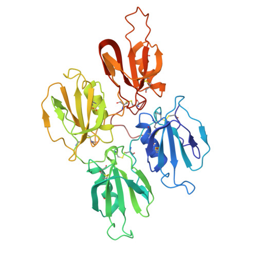
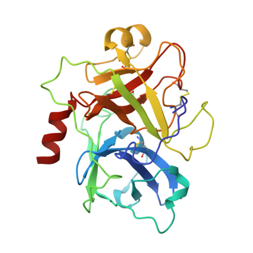
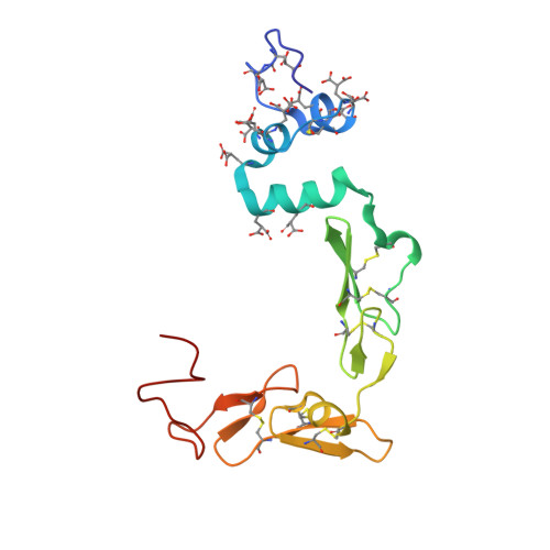
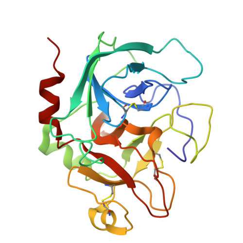
    Factor XI (FXI) occupies a unique and clinically significant niche, bridging the tissue factor-driven and contact-driven coagulation pathways. Irrespective of the trigger, activated factor XI (FXIa) contributes to clotting by activating factor IX (FIX). Biochemical studies established that this reaction requires the membrane-binding FIX-Gla domain to engage an exosite on the FXIa Apple 3 (A3) domain, that only become available upon FXI activation. Structural data for FXIa, FIX, FIXaβ, and the FXIa:FIX complex are lacking; current understanding relies on zymogen FXI crystal structures and homology modeling of FXIa after kallikrein. Elucidate the high-resolution structure of FXIa in functionally relevant conformation in complex with FIX. We utilized cryogenic electron microscopy (cryo-EM) to determine the structures of human FXIa in complex with its full-length substrate, FIX, and activated product, FIXaβ. We report the first cryo-EM structures of FXIa in complex with its substrate, FIX, and activated FIX (FIXaβ). The structures capture a functionally relevant conformational change in the FXIa catalytic domain and reveals the first view of the entire FIX and FIXaβ. Critically, we visualize the FIX-Gla domain precisely docked to the FXIa-A3 exosite on both subunits of the FXIa dimer. We also define the first step of proteolysis, visualizing the FIX Arg145 inserted into the primary specificity pocket of FXIa. The structures define the full FXIa:FIX interface providing a structural template for understanding the sequential activation of FIX and for developing a new class of selective allosteric antithrombotic agents.


  • Organizational Affiliation
    • Edward A. Doisy Department of Biochemistry and Molecular Biology, Saint Louis University School of Medicine, St. Louis, MO 63104 USA. Electronic address: Bassem.mohammed@health.slu.edu.

Macromolecule Content 

  • Total Structure Weight: 112.38 kDa 
  • Atom Count: 7,841 
  • Modeled Residue Count: 985 
  • Deposited Residue Count: 985 
  • Unique protein chains: 4

Macromolecules

Find similar proteins by:|  3D Structure
Entity ID: 1
MoleculeChains  Sequence LengthOrganismDetailsImage
Coagulation factor XIa heavy chain369Homo sapiensMutation(s): 0 
Gene Names: F11
EC: 3.4.21.27
UniProt & NIH Common Fund Data Resources
Find proteins for P03951 (Homo sapiens)
Explore P03951 
Go to UniProtKB:  P03951
PHAROS:  P03951
GTEx:  ENSG00000088926 
Entity Groups
Sequence Clusters30% Identity50% Identity70% Identity90% Identity95% Identity100% Identity
UniProt GroupP03951
Glycosylation
Glycosylation Sites: 2Go to GlyGen: P03951-1
Sequence Annotations
Expand
Reference Sequence
Find similar proteins by:|  3D Structure
Entity ID: 2
MoleculeChains  Sequence LengthOrganismDetailsImage
Coagulation factor XIa light chain236Homo sapiensMutation(s): 1 
Gene Names: F11
EC: 3.4.21.27
UniProt & NIH Common Fund Data Resources
Find proteins for P03951 (Homo sapiens)
Explore P03951 
Go to UniProtKB:  P03951
PHAROS:  P03951
GTEx:  ENSG00000088926 
Entity Groups
Sequence Clusters30% Identity50% Identity70% Identity90% Identity95% Identity100% Identity
UniProt GroupP03951
Glycosylation
Glycosylation Sites: 2Go to GlyGen: P03951-1
Sequence Annotations
Expand
Reference Sequence
Find similar proteins by:|  3D Structure
Entity ID: 3
MoleculeChains  Sequence LengthOrganismDetailsImage
Coagulation factor IXa light chain145Homo sapiensMutation(s): 0 
EC: 3.4.21.22
UniProt & NIH Common Fund Data Resources
Find proteins for P00740 (Homo sapiens)
Explore P00740 
Go to UniProtKB:  P00740
PHAROS:  P00740
GTEx:  ENSG00000101981 
Entity Groups
Sequence Clusters30% Identity50% Identity70% Identity90% Identity95% Identity100% Identity
UniProt GroupP00740
Sequence Annotations
Expand
Reference Sequence
Find similar proteins by:|  3D Structure
Entity ID: 4
MoleculeChains  Sequence LengthOrganismDetailsImage
Coagulation factor IXa heavy chain235Homo sapiensMutation(s): 0 
EC: 3.4.21.22
UniProt & NIH Common Fund Data Resources
Find proteins for P00740 (Homo sapiens)
Explore P00740 
Go to UniProtKB:  P00740
PHAROS:  P00740
GTEx:  ENSG00000101981 
Entity Groups
Sequence Clusters30% Identity50% Identity70% Identity90% Identity95% Identity100% Identity
UniProt GroupP00740
Sequence Annotations
Expand
Reference Sequence

Small Molecules

Ligands 2 Unique
IDChains Name / Formula / InChI Key2D Diagram3D Interactions
NAG
(Subject of Investigation/LOI)

Query on NAG


Download:Ideal Coordinates CCD File
E [auth A],
F [auth A],
G [auth B],
H [auth B]
2-acetamido-2-deoxy-beta-D-glucopyranose
C8 H15 N O6
OVRNDRQMDRJTHS-FMDGEEDCSA-N
CA
(Subject of Investigation/LOI)

Query on CA


Download:Ideal Coordinates CCD File
I [auth C]
J [auth C]
K [auth C]
L [auth C]
M [auth C]
I [auth C],
J [auth C],
K [auth C],
L [auth C],
M [auth C],
N [auth C],
O [auth C],
P [auth C]
CALCIUM ION
Ca
BHPQYMZQTOCNFJ-UHFFFAOYSA-N
Modified Residues  1 Unique
IDChains TypeFormula2D DiagramParent
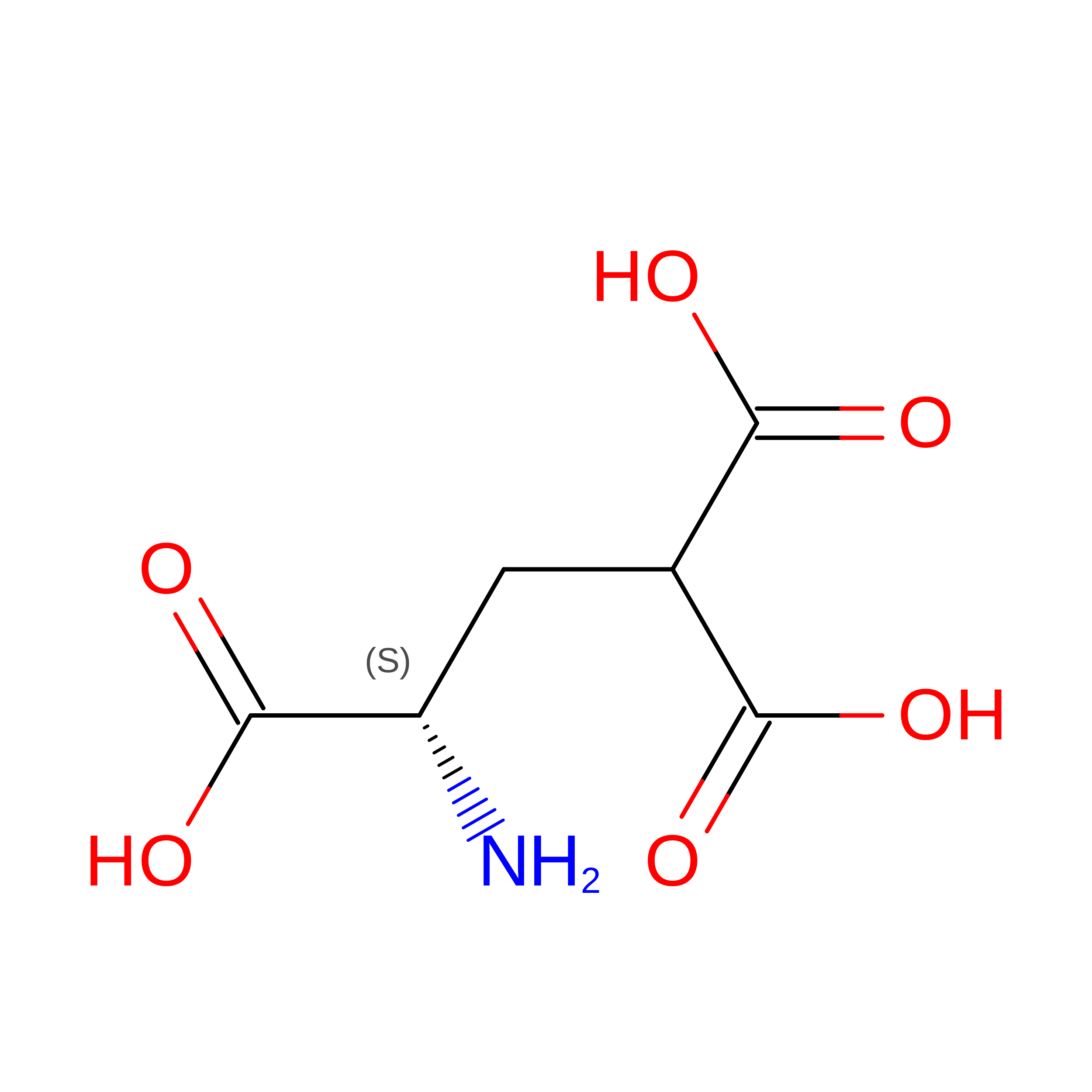
CGU
Query on CGU
C
L-PEPTIDE LINKINGC6 H9 N O6GLU

Experimental Data & Validation

Experimental Data

  • Method: ELECTRON MICROSCOPY
  • Resolution: 3.40 Å
  • Resolution: 3.40 Å
  • Resolution: 3.40 Å
  • Resolution: 3.40 Å
  • Aggregation State: PARTICLE 
  • Reconstruction Method: SINGLE PARTICLE 
EM Software:
TaskSoftware PackageVersion
RECONSTRUCTIONcryoSPARC4.7.1
MODEL REFINEMENTPHENIX1.21.2_5419
MODEL REFINEMENTCoot0.9.8.95

Structure Validation

View Full Validation Report


Entry History 

& Funding Information

Deposition Data


Funding OrganizationLocationGrant Number
Other privateUnited StatesDoisy Fund of the Edward A. Doisy Department of Biochemistry and Molecular Biology
Childrens Discovery Institute of Washington University and St. Louis Childrens HospitalUnited StatesCDI-CORE-2015-505 and CDI-CORE-2019-813
The Foundation for Barnes-Jewish HospitalUnited States3770
National Institutes of Health/National Institute of Diabetes and Digestive and Kidney Disease (NIH/NIDDK)United StatesDK020579
National Institutes of Health/National Cancer Institute (NIH/NCI)United StatesCA091842

Revision History  (Full details and data files)

  • Version 1.0: 2026-09-09
    Type: Initial release