9ZTB | pdb_00009ztb

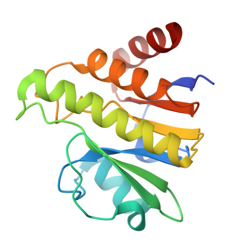
Crystal Structure of the macrodomain in the complex with suramin analog from Venezuelan equine encephalitis virus


Experimental Data Snapshot

  • Method: X-RAY DIFFRACTION
  • Resolution: 1.67 Å
  • R-Value Free: 
    0.252 (Depositor), 0.253 (DCC) 
  • R-Value Work: 
    0.203 (Depositor), 0.203 (DCC) 
  • R-Value Observed: 
    0.206 (Depositor) 

Starting Model: in silico
View more details

wwPDB Validation 3D Report Full Report

Validation slider image for 9ZTB

Ligand Structure Quality Assessment 


This is version 1.0 of the entry. See complete history

Literature

Crystal Structure of the macrodomain in the complex with suramin analog from Venezuelan equine encephalitis virus

Kim, Y.Stols, L.Endres, M.Babnigg, G.Gora, N.Hura, G.Joachimiak, A.Center for Structural Biology of Infectious Diseases (CSBID)

To be published.

Macromolecule Content 

  • Total Structure Weight: 36.39 kDa 
  • Atom Count: 2,732 
  • Modeled Residue Count: 313 
  • Deposited Residue Count: 326 
  • Unique protein chains: 1

Macromolecules

Find similar proteins by:|  3D Structure
Entity ID: 1
MoleculeChains  Sequence LengthOrganismDetailsImage
Polyprotein P1234
A, B
163Venezuelan equine encephalitis virusMutation(s): 0 
Gene Names: NSP
Entity Groups
Sequence Clusters30% Identity50% Identity70% Identity90% Identity95% Identity100% Identity
Sequence Annotations
Expand
Reference Sequence

Small Molecules

Ligands 1 Unique
IDChains Name / Formula / InChI Key2D Diagram3D Interactions
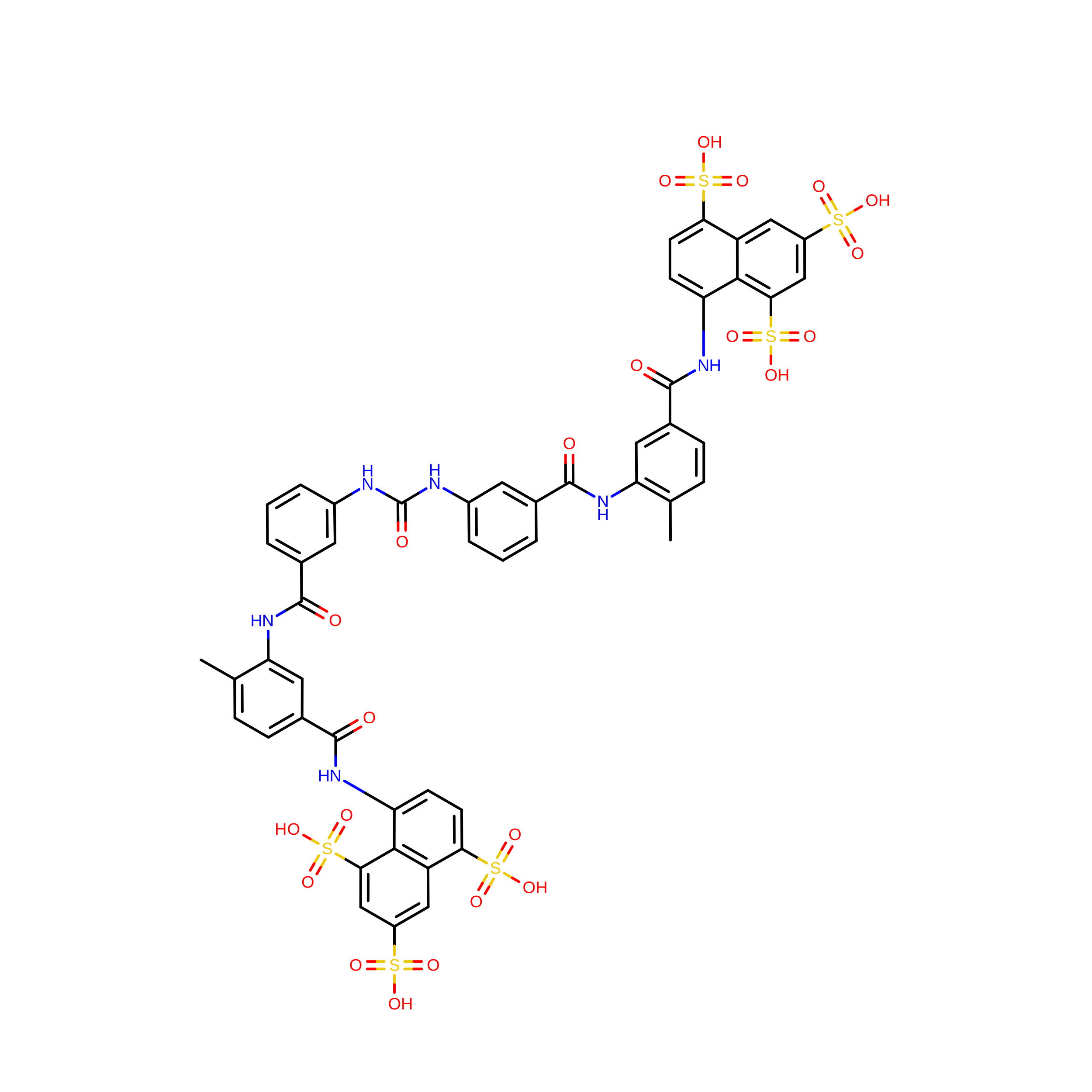
SVR
(Subject of Investigation/LOI)

Query on SVR


Download:Ideal Coordinates CCD File
C [auth A]8,8'-[CARBONYLBIS[IMINO-3,1-PHENYLENECARBONYLIMINO(4-METHYL-3,1-PHENYLENE)CARBONYLIMINO]]BIS-1,3,5-NAPHTHALENETRISULFON IC ACID
C51 H40 N6 O23 S6
FIAFUQMPZJWCLV-UHFFFAOYSA-N

Experimental Data & Validation

Experimental Data

  • Method: X-RAY DIFFRACTION
  • Resolution: 1.67 Å
  • R-Value Free:  0.252 (Depositor), 0.253 (DCC) 
  • R-Value Work:  0.203 (Depositor), 0.203 (DCC) 
  • R-Value Observed: 0.206 (Depositor) 
Space Group: P 1
Unit Cell:
Length ( Å )Angle ( ˚ )
a = 39.471α = 82.633
b = 39.65β = 69.389
c = 56.06γ = 61.974
Software Package:
Software NamePurpose
PHENIXrefinement
HKL-3000data reduction
HKL-3000data scaling
HKL-3000phasing
MOLREPphasing

Structure Validation

View Full Validation Report


Ligand Structure Quality Assessment 


Entry History 

& Funding Information

Deposition Data


Funding OrganizationLocationGrant Number
National Institutes of Health/National Institute Of Allergy and Infectious Diseases (NIH/NIAID)United States--

Revision History  (Full details and data files)

  • Version 1.0: 2026-08-12
    Type: Initial release