9ZGF | pdb_00009zgf

The complex of HSV-1 proteins UL9 and ICP8 with forked DNA


Experimental Data Snapshot

  • Method: ELECTRON MICROSCOPY
  • Resolution: 3.10 Å
  • Aggregation State: PARTICLE 
  • Reconstruction Method: SINGLE PARTICLE 

wwPDB Validation 3D Report Full Report

Validation slider image for 9ZGF

This is version 1.1 of the entry. See complete history

Literature

Structural insights into HSV-1 origin unwinding by the viral proteins UL9 and ICP8.

Baranovskiy, A.G.Morstadt, L.M.Romero, E.E.Babayeva, N.D.Tahirov, T.H.

(2026) Nucleic Acids Res 54

  • DOI: https://doi.org/10.1093/nar/gkag580
  • Primary Citation Related Structures: 
    9ZGF

  • PubMed Abstract: 

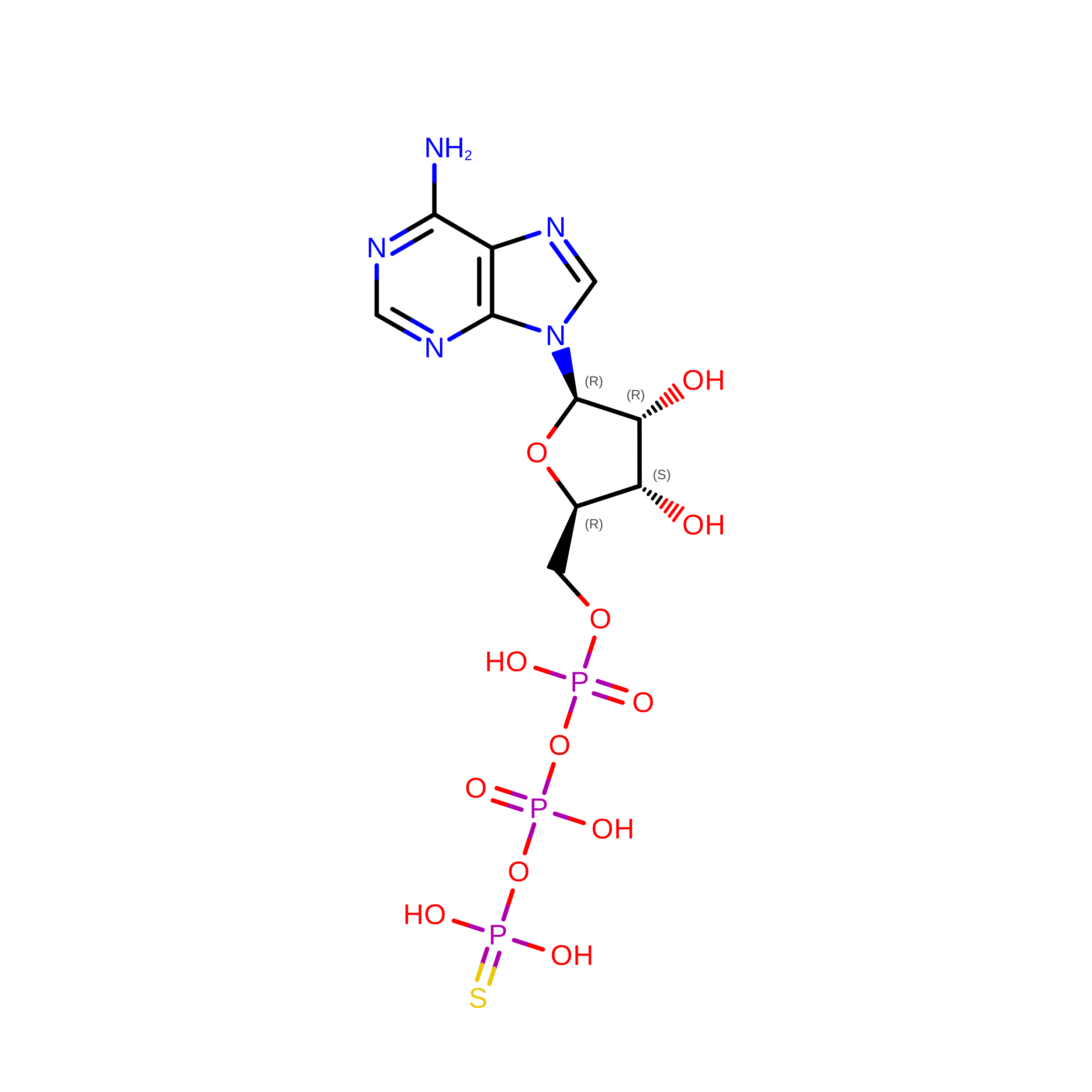
    Herpes simplex virus type 1 (HSV-1) causes lifelong infections in human cells and is associated with a range of diseases. HSV-1 DNA replication requires seven viral proteins, including the major DNA-binding protein ICP8, the origin-binding protein UL9, and proteins that comprise the helicase-primase and DNA polymerase complexes. UL9 functions as a DNA helicase that specifically recognizes and binds to the viral origins of replication, OriS and OriL. Here we report the cryo-EM structure of the UL9/ICP8/DNA/ATPγS complex at an overall resolution of 3.18 Å. This structure revealed that UL9 employs an α-helix to separate the DNA strands and captures the initial step of OriS unwinding, in which the C-terminal domain of UL9 specifically binds to the major groove of a DNA double helix, while the N-terminal helicase domain engages the unwound leading and lagging strands. ICP8 interacts with the extreme C-terminal region of UL9, preventing UL9 dimerization. Simultaneously, it binds and stabilizes the leading-strand DNA adjacent to UL9. Together, these findings provide mechanistic insight into UL9-driven DNA unwinding and the cooperative action of UL9 and ICP8 at HSV-1 replication origins, establishing a structural framework for the rational interpretation of prior biochemical data and for the design of new antiviral drugs.


  • Organizational Affiliation
    • Eppley Institute for Research in Cancer and Allied Diseases, Fred & Pamela Buffett Cancer Center, University of Nebraska Medical Center, Omaha, NE 68198, United States.

Macromolecule Content 

  • Total Structure Weight: 245.24 kDa 
  • Atom Count: 15,403 
  • Modeled Residue Count: 1,896 
  • Deposited Residue Count: 2,118 
  • Unique protein chains: 2
  • Unique nucleic acid chains: 2

Macromolecules


Find similar proteins by:|  3D Structure
Entity ID: 1
MoleculeChains  Sequence LengthOrganismDetailsImage
Major DNA-binding protein1,196Human alphaherpesvirus 1 strain 17Mutation(s): 0 
Gene Names: DBPICP8UL29
UniProt
Find proteins for P04296 (Human herpesvirus 1 (strain 17))
Explore P04296 
Go to UniProtKB:  P04296
Entity Groups
Sequence Clusters30% Identity50% Identity70% Identity90% Identity95% Identity100% Identity
UniProt GroupP04296
Sequence Annotations
Expand
Reference Sequence
Find similar proteins by:|  3D Structure
Entity ID: 2
MoleculeChains  Sequence LengthOrganismDetailsImage
Replication origin-binding proteinB [auth C]851Human alphaherpesvirus 1 strain 17Mutation(s): 0 
Gene Names: UL9
UniProt
Find proteins for P10193 (Human herpesvirus 1 (strain 17))
Explore P10193 
Go to UniProtKB:  P10193
Entity Groups
Sequence Clusters30% Identity50% Identity70% Identity90% Identity95% Identity100% Identity
UniProt GroupP10193
Sequence Annotations
Expand
Reference Sequence
Find similar nucleic acids by:  (by identity cutoff) 
Entity ID: 3
MoleculeChains LengthOrganismImage
DNA (40-MER)C [auth D]40Human alphaherpesvirus 1 strain 17
Sequence Annotations
Expand
Reference Sequence
Find similar nucleic acids by:  (by identity cutoff) 
Entity ID: 4
MoleculeChains LengthOrganismImage
DNA (31-MER)D [auth E]31Human alphaherpesvirus 1 strain 17
Sequence Annotations
Expand
Reference Sequence

Experimental Data & Validation

Experimental Data

  • Method: ELECTRON MICROSCOPY
  • Resolution: 3.10 Å
  • Aggregation State: PARTICLE 
  • Reconstruction Method: SINGLE PARTICLE 
EM Software:
TaskSoftware PackageVersion
MODEL REFINEMENTPHENIX1.21.2_5419:
RECONSTRUCTIONcryoSPARC

Structure Validation

View Full Validation Report


Entry History 

& Funding Information

Deposition Data


Funding OrganizationLocationGrant Number
National Institutes of Health/National Institute of General Medical Sciences (NIH/NIGMS)United StatesR35 GM152032

Revision History  (Full details and data files)

  • Version 1.0: 2026-06-10
    Type: Initial release
  • Version 1.1: 2026-06-24
    Changes: Data collection, Database references