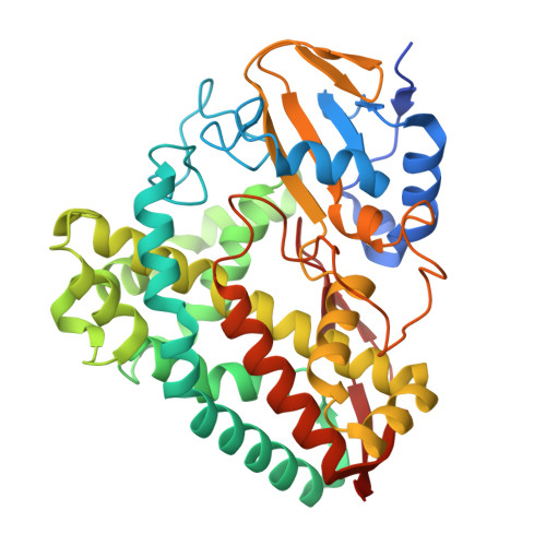
Structural basis for substrate-dependent allostery in oxygen activation by a cytochrome P450 enzyme revealed by analysis at different temperatures.
Podgorski, M.N., McDougal, D.P., Campbell, E.C., Bruning, J.B., Bell, S.G.(2026) Chem Sci 17: 7742-7755
- PubMed: 41767801 Search on PubMedSearch on PubMed Central
- DOI: https://doi.org/10.1039/d5sc07539d
- Primary Citation Related Structures:
9DOE, 9MIM, 9MIO, 9MJE, 9MJF, 9MJJ, 9MJK, 9PLS, 9PMA, 9PMC, 9ZEG, 9ZEH, 9ZEI - PubMed Abstract:
Cytochrome P450 (CYP) enzymes are ubiquitous and important monooxygenases whose archetypal reaction is to insert an oxygen atom from dioxygen into unactivated carbon-hydrogen bonds. They require the orchestrated delivery of electrons as well as protons from the solvent. The latter is controlled through an "acid-alcohol pair" of residues located above the heme though the precise details of proton delivery are unresolved. Here, using variable-temperature X-ray crystallography and all-atom molecular dynamics simulations of the bacterial CYP199A4 enzyme we demonstrate that the conformation of the acidic residue D251, of the "acid-alcohol" pair is allosterically coupled to the heme and the substrate. In general, and in common with other CYPs, the side chain of D251 favours the 'out' of the active site orientation. In this enzyme this overcomes incompatibility with hydrophobic residues. This side chain can rotate into the active site, and this is allosterically coupled to the presence of a distal heme ligand and other structural changes at the E-helix, C-terminal loop and on the proximal side of the heme. These and other structural changes can be related to differences in water molecule access to and egress from the distal side of the heme, which would facilitate proton delivery during the catalytic cycle. Comparison of the different environments of the side chain of D251 in CYP199A4 with the equivalent acidic residue in other diverse CYP enzymes suggest that there may not be a 'universal' model for proton transfer in CYP enzymes, but that allosteric effects and transient interactions are critically important.
- Department of Chemistry, University of Adelaide Adelaide South Australia 5005 Australia stephen.bell@adelaide.edu.au.
Organizational Affiliation: