9ZD5 | pdb_00009zd5

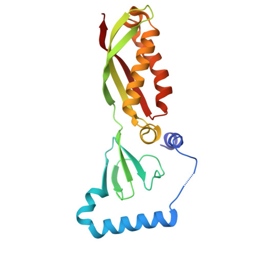
Human Stomatin - intramembrane region


Experimental Data Snapshot

  • Method: ELECTRON MICROSCOPY
  • Resolution: 2.20 Å
  • Aggregation State: PARTICLE 
  • Reconstruction Method: SINGLE PARTICLE 

wwPDB Validation 3D Report Full Report

Validation slider image for 9ZD5

This is version 1.1 of the entry. See complete history

Literature

Stomatin encapsulates aquaporin-1 and urea transporter-B in the erythrocyte membrane.

Vallese, F.Li, H.Barazzuol, L.Cali, T.Clarke, O.B.

(2026) Sci Adv 12: eaec1721-eaec1721

  • DOI: https://doi.org/10.1126/sciadv.aec1721
  • Primary Citation Related Structures: 
    9Z7U, 9ZCZ, 9ZD0, 9ZD2, 9ZD5

  • PubMed Abstract: 

    Stomatin is a ubiquitous and highly expressed protein in erythrocytes, which associates with cholesterol-rich microdomains in the plasma membrane and is known to regulate the activity of multiple ion channels and transporters, but the structural basis of association with stomatin targets remains unknown. Here, we describe high-resolution structures of multiple stomatin complexes with endogenous binding partners isolated from human erythrocyte membranes, revealing that stomatin specifically associates with two membrane proteins involved in water transport and cell volume regulation, aquaporin-1 and the urea transporter SLC14A1. Together, our results reveal the structural basis of stomatin oligomerization, membrane association, and target recruitment and identify a putative role for stomatin in the regulation of osmotic balance in the erythrocyte.


  • Organizational Affiliation
    • Structural Biology Initiative, CUNY Advanced Science Research Center, New York, NY 10031, USA.

Macromolecule Content 

  • Total Structure Weight: 91.6 kDa 
  • Atom Count: 6,340 
  • Modeled Residue Count: 776 
  • Deposited Residue Count: 776 
  • Unique protein chains: 1

Macromolecules

Find similar proteins by:|  3D Structure
Entity ID: 1
MoleculeChains  Sequence LengthOrganismDetailsImage
StomatinA [auth I],
B [auth E],
C [auth G],
D [auth H]
194Homo sapiensMutation(s): 0 
UniProt & NIH Common Fund Data Resources
Find proteins for P27105 (Homo sapiens)
Explore P27105 
Go to UniProtKB:  P27105
PHAROS:  P27105
GTEx:  ENSG00000148175 
Entity Groups
Sequence Clusters30% Identity50% Identity70% Identity90% Identity95% Identity100% Identity
UniProt GroupP27105
Sequence Annotations
Expand
Reference Sequence

Small Molecules

Ligands 3 Unique
IDChains Name / Formula / InChI Key2D Diagram3D Interactions
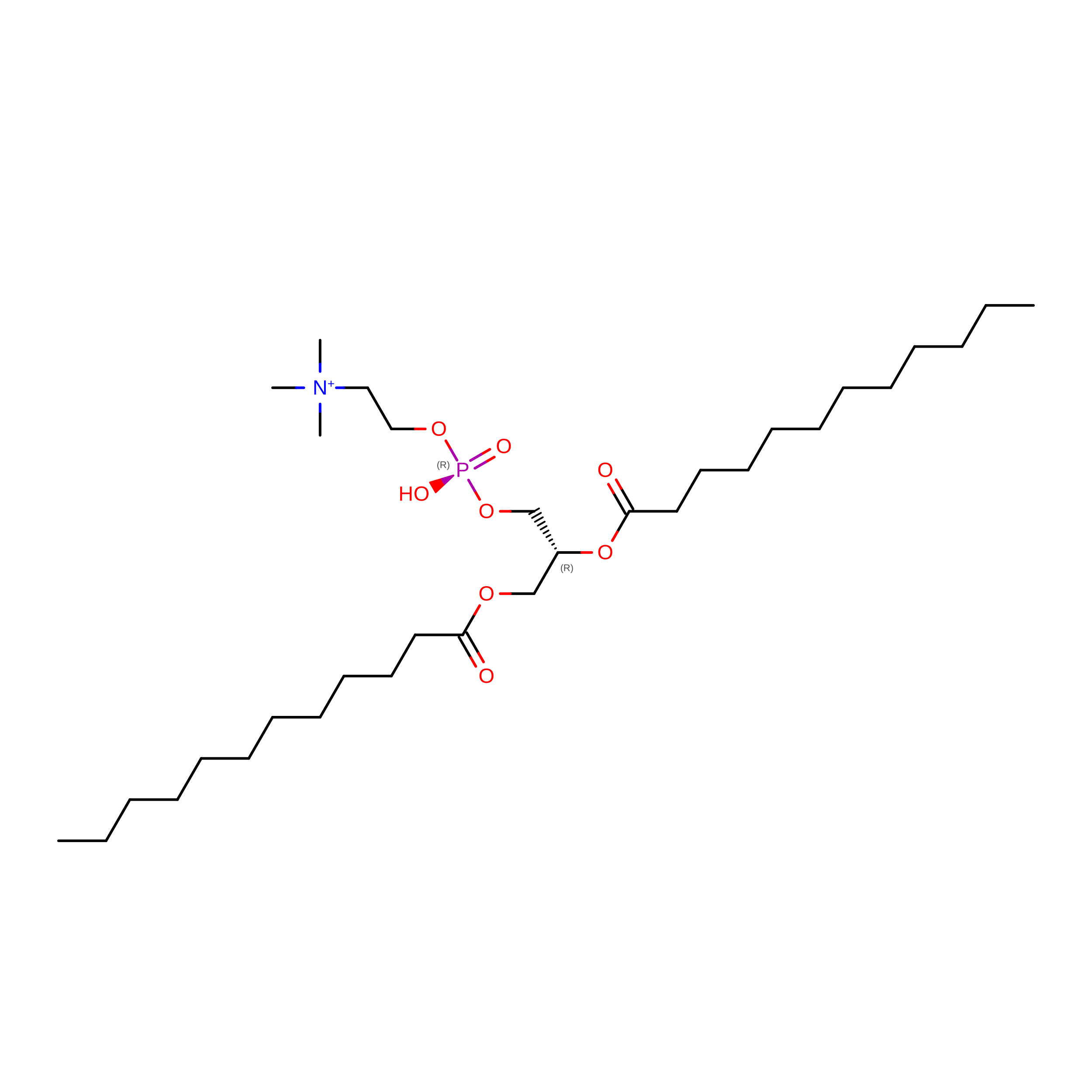
PLC
(Subject of Investigation/LOI)

Query on PLC


Download:Ideal Coordinates CCD File
H [auth I],
M [auth E],
R [auth H],
S [auth H]
DIUNDECYL PHOSPHATIDYL CHOLINE
C32 H65 N O8 P
IJFVSSZAOYLHEE-SSEXGKCCSA-O
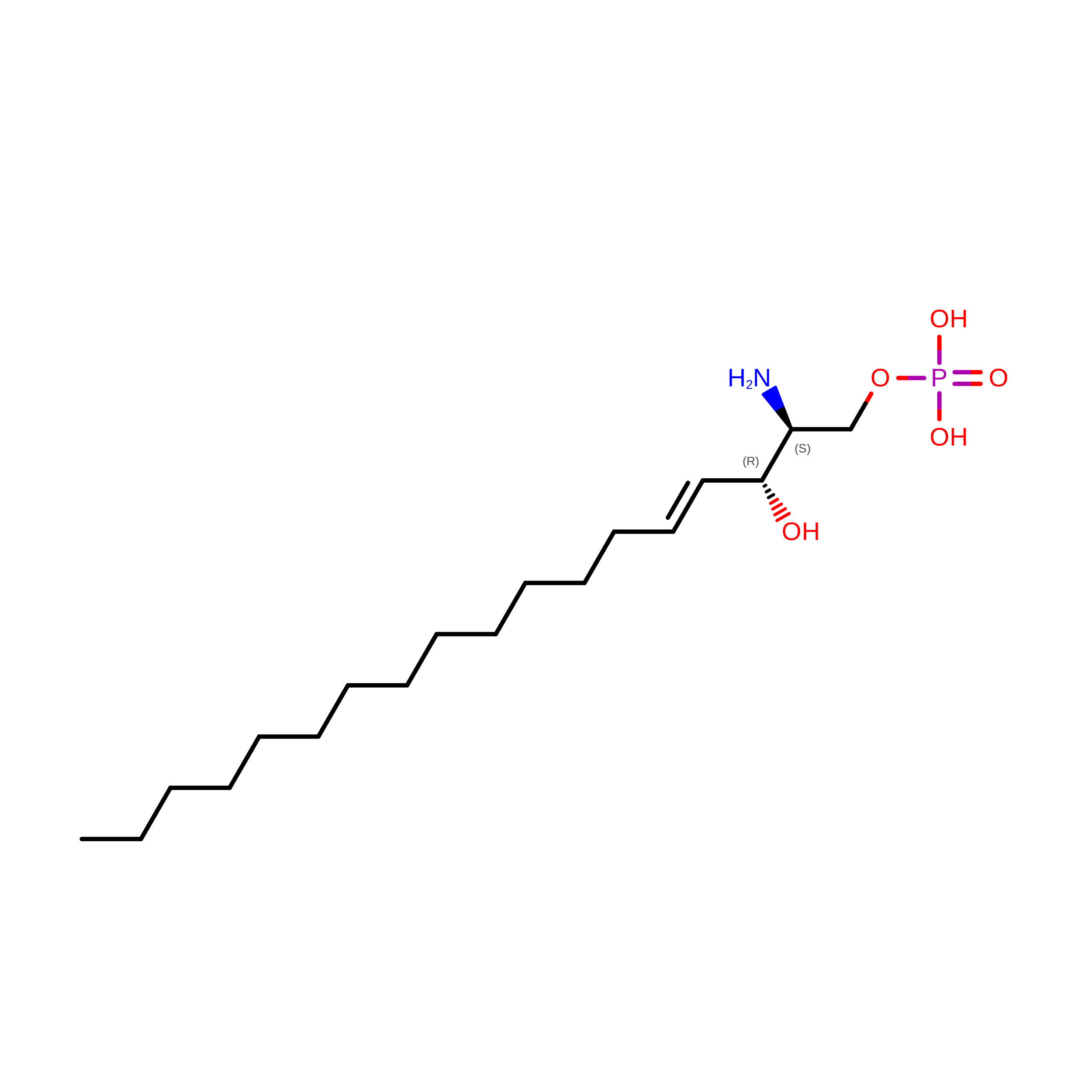
S1P
(Subject of Investigation/LOI)

Query on S1P


Download:Ideal Coordinates CCD File
I,
N [auth E],
W [auth H],
X [auth H]
(2S,3R,4E)-2-amino-3-hydroxyoctadec-4-en-1-yl dihydrogen phosphate
C18 H38 N O5 P
DUYSYHSSBDVJSM-KRWOKUGFSA-N
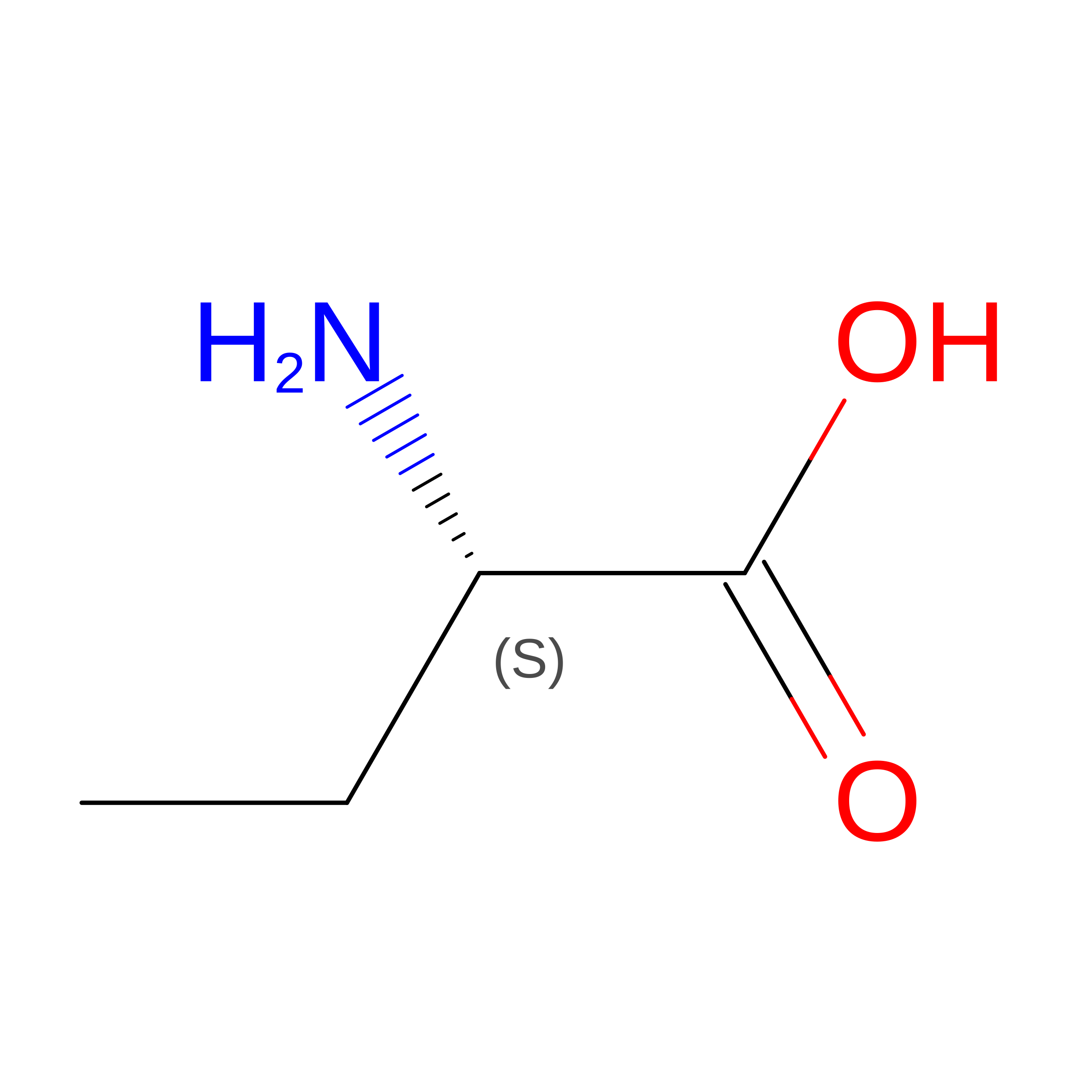
PLM

Query on PLM


Download:Ideal Coordinates CCD File
E [auth I]
F [auth I]
G [auth I]
J [auth E]
K [auth E]
E [auth I],
F [auth I],
G [auth I],
J [auth E],
K [auth E],
L [auth E],
O [auth G],
P [auth G],
Q [auth G],
T [auth H],
U [auth H],
V [auth H]
PALMITIC ACID
C16 H32 O2
IPCSVZSSVZVIGE-UHFFFAOYSA-N

Experimental Data & Validation

Experimental Data

  • Method: ELECTRON MICROSCOPY
  • Resolution: 2.20 Å
  • Aggregation State: PARTICLE 
  • Reconstruction Method: SINGLE PARTICLE 
EM Software:
TaskSoftware PackageVersion
RECONSTRUCTIONcryoSPARC

Structure Validation

View Full Validation Report


Entry History 

& Funding Information

Deposition Data


Funding OrganizationLocationGrant Number
Not funded--

Revision History  (Full details and data files)

  • Version 1.0: 2026-03-04
    Type: Initial release
  • Version 1.1: 2026-04-29
    Changes: Data collection, Database references