9ZBL | pdb_00009zbl

Helical Reconstruction of the Human Cardiac F-Actin-Tropomyosin Complex


Experimental Data Snapshot

  • Method: ELECTRON MICROSCOPY
  • Resolution: 2.79 Å
  • Aggregation State: FILAMENT 
  • Reconstruction Method: HELICAL 

Starting Model: experimental
View more details

wwPDB Validation 3D Report Full Report

Validation slider image for 9ZBL

This is version 1.0 of the entry. See complete history. 

Literature

Pseudo-acetylation of ACTC1 K326 and K328 promotes dysinhibition of reconstituted human cardiac thin filaments.

Chitre, K., Karpicheva, O.E., King, C.J., Rynkiewicz, M.J., Fenwick, A.J., Dawson, J.F., Foster, D.B., Lehman, W., Cammarato, A.

(2025) J Mol Cell Cardiol 212: 10-15

  • DOI: https://doi.org/10.1016/j.yjmcc.2025.12.008
  • Primary Citation Related Structures: 
    9ZBL, 9ZBP

  • PubMed Abstract: 

    Electrostatic interactions between actin residues K326 and K328 and tropomyosin bias tropomyosin to an F-actin location where it blocks myosin attachment. K326/328 acetylation neutralizes their charge, potentially disrupting thin filament-based contractile regulation. We verified acetylation of K326/328 on human cardiac actin (ACTC1) and generated recombinant K326/328Q, pseudo-acetylated ACTC1. Pseudo-acetylation reduced inhibition of myosin-driven motility of F-actin-tropomyosin and F-actin-tropomyosin-troponin at low Ca 2+ . Cryo-EM-based and computational modeling revealed that pseudo-acetylation did not alter tropomyosin positioning along F-actin but decreased local F-actin-tropomyosin interaction energy. Thus, by reducing the energetic demands required for myosin to displace tropomyosin, ACTC1 K326/328 acetylation may promote contractile activation.


  • Organizational Affiliation: 
    • Department of Medicine, Division of Cardiology, Johns Hopkins University, 720 Rutland Avenue, Baltimore, MD 21205, USA.

Macromolecule Content 

  • Total Structure Weight: 386.79 kDa 
  • Atom Count: 21,570 
  • Modeled Residue Count: 2,722 
  • Deposited Residue Count: 3,406 
  • Unique protein chains: 2

Macromolecules

Find similar proteins by:|  3D Structure
Entity ID: 1
MoleculeChains  Sequence LengthOrganismDetailsImage
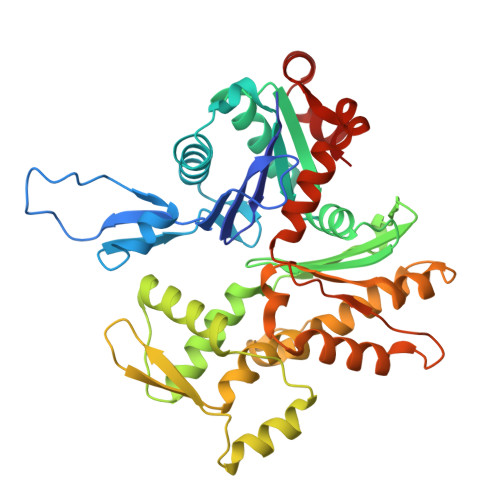
Actin, alpha cardiac muscle 1
A, B, C, D, E
A, B, C, D, E, F
377Homo sapiensMutation(s): 0 
Gene Names: ACTC1, ACTC
EC: 3.6.4
UniProt & NIH Common Fund Data Resources
Find proteins for P68032 (Homo sapiens)
Explore P68032 
Go to UniProtKB:  P68032
PHAROS:  P68032
GTEx:  ENSG00000159251 
Entity Groups
Sequence Clusters30% Identity50% Identity70% Identity90% Identity95% Identity100% Identity
UniProt GroupP68032
Sequence Annotations
Expand
Reference Sequence
Find similar proteins by:|  3D Structure
Entity ID: 2
MoleculeChains  Sequence LengthOrganismDetailsImage
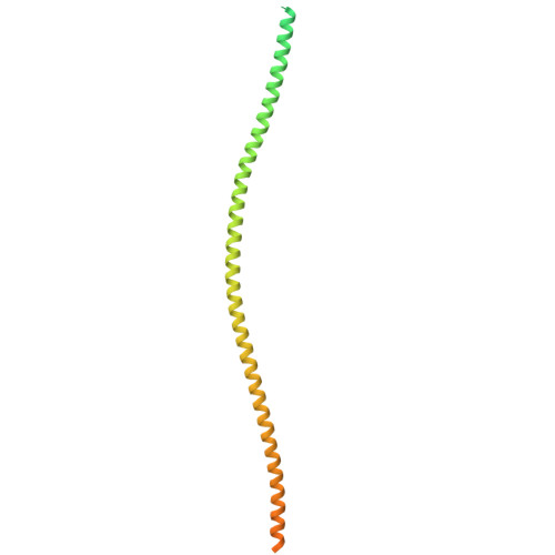
Tropomyosin alpha-1 chainG [auth M],
H [auth N],
I [auth O],
J [auth P]
286Homo sapiensMutation(s): 0 
Gene Names: TPM1, C15orf13, TMSA
UniProt & NIH Common Fund Data Resources
Find proteins for P09493 (Homo sapiens)
Explore P09493 
Go to UniProtKB:  P09493
PHAROS:  P09493
GTEx:  ENSG00000140416 
Entity Groups
Sequence Clusters30% Identity50% Identity70% Identity90% Identity95% Identity100% Identity
UniProt GroupP09493
Sequence Annotations
Expand
Reference Sequence

Small Molecules

Ligands 2 Unique
IDChains Name / Formula / InChI Key2D Diagram3D Interactions
ADP

Query on ADP


Download:Ideal Coordinates CCD File
K [auth A]
M [auth B]
O [auth C]
Q [auth D]
S [auth E]
K [auth A],
M [auth B],
O [auth C],
Q [auth D],
S [auth E],
U [auth F]
ADENOSINE-5'-DIPHOSPHATE
C10 H15 N5 O10 P2
XTWYTFMLZFPYCI-KQYNXXCUSA-N
MG

Query on MG


Download:Ideal Coordinates CCD File
L [auth A]
N [auth B]
P [auth C]
R [auth D]
T [auth E]
L [auth A],
N [auth B],
P [auth C],
R [auth D],
T [auth E],
V [auth F]
MAGNESIUM ION
Mg
JLVVSXFLKOJNIY-UHFFFAOYSA-N

Experimental Data & Validation

Experimental Data

  • Method: ELECTRON MICROSCOPY
  • Resolution: 2.79 Å
  • Aggregation State: FILAMENT 
  • Reconstruction Method: HELICAL 
EM Software:
TaskSoftware PackageVersion
RECONSTRUCTIONcryoSPARCv4.5.3
MODEL REFINEMENTPHENIX2.0.5867

Structure Validation

View Full Validation Report


Entry History 

& Funding Information

Deposition Data


Funding OrganizationLocationGrant Number
National Institutes of Health/National Heart, Lung, and Blood Institute (NIH/NHLBI)United StatesHL036153
National Institutes of Health/National Heart, Lung, and Blood Institute (NIH/NHLBI)United StatesHL124091

Revision History  (Full details and data files)

  • Version 1.0: 2026-01-14
    Type: Initial release