9Z9B | pdb_00009z9b

Gated state sheep connexin-50 in DMPC nanodiscs at neutral pH


Experimental Data Snapshot

  • Method: ELECTRON MICROSCOPY
  • Resolution: 2.70 Å
  • Aggregation State: PARTICLE 
  • Reconstruction Method: SINGLE PARTICLE 

Starting Model: experimental
View more details

wwPDB Validation   3D Report Full Report


This is version 1.0 of the entry. See complete history


Literature

Reversible lipid-mediated pH-gating of connexin-46/50 by cryo-EM.

Jarodsky, J.M.Myers, J.B.Reichow, S.L.

(2026) Nat Commun 

  • DOI: https://doi.org/10.1038/s41467-026-68311-9
  • Primary Citation of Related Structures:  
    9Z7P, 9Z7W, 9Z81, 9Z82, 9Z8F, 9Z8L, 9Z8M, 9Z9B, 9Z9G, 9Z9H, 9Z9S, 9Z9W, 9Z9X, 9Z9Y, 9ZA3, 9ZA4

  • PubMed Abstract: 

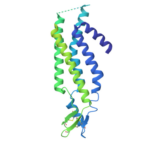
    Gap junctions, formed by connexin proteins, establish direct electrical and metabolic coupling between cells, enabling coordinated tissue responses. These channels universally respond to intracellular pH changes, closing under acidic conditions to limit the spread of cytotoxic signals during cellular stress, such as ischemia. Using cryo-electron microscopy (cryo-EM), we uncover insights into the structural mechanism of pH-gating in native lens connexin-46/50 (Cx46/50) gap junctions. Mild acidification drives lipid infiltration into the channel pore, displacing the N-terminal (NT) domain and stabilizing pore closure. Lipid involvement is shown to be both essential and fully reversible. Structural transitions involve an ensemble of gated states formed through non-cooperative NT domain movement as well as minor populations of a distinct destabilized open-state. These findings provide molecular insights into pH-gating dynamics, illustrating how structural changes may regulate gap junction function under cellular stress and linking Cx46/50 dysregulation to age-related cataract formation.


  • Organizational Affiliation
    • Department of Chemical Physiology and Biochemistry, Oregon Health and Science University, Portland, OR, USA.

Macromolecules
Find similar proteins by:  (by identity cutoff)  |  3D Structure
Entity ID: 1
MoleculeChains Sequence LengthOrganismDetailsImage
Gap junction alpha-8 protein
A, B, C, D, E
A, B, C, D, E, F, G, H, I, J, K, L
440Ovis ariesMutation(s): 0 
UniProt
Find proteins for P55917 (Ovis aries)
Explore P55917 
Go to UniProtKB:  P55917
Entity Groups  
Sequence Clusters30% Identity50% Identity70% Identity90% Identity95% Identity100% Identity
UniProt GroupP55917
Sequence Annotations
Expand
  • Reference Sequence
Small Molecules
Ligands 1 Unique
IDChains Name / Formula / InChI Key2D Diagram3D Interactions
MC3
Query on MC3

Download Ideal Coordinates CCD File 
AA [auth A]
AB [auth C]
AC [auth E]
AD [auth F]
AE [auth H]
AA [auth A],
AB [auth C],
AC [auth E],
AD [auth F],
AE [auth H],
AF [auth I],
AG [auth K],
BA [auth A],
BB [auth C],
BC [auth E],
BD [auth F],
BE [auth H],
BF [auth J],
BG [auth K],
CA [auth B],
CB [auth C],
CC [auth E],
CD [auth F],
CE [auth H],
CF [auth J],
CG [auth K],
DA [auth B],
DB [auth C],
DC [auth E],
DD [auth F],
DE [auth H],
DF [auth J],
DG [auth K],
EA [auth B],
EB [auth C],
EC [auth E],
ED [auth G],
EE [auth H],
EF [auth J],
EG [auth K],
FA [auth B],
FB [auth C],
FC [auth E],
FD [auth G],
FE [auth H],
FF [auth J],
FG [auth K],
GA [auth B],
GB [auth C],
GC [auth E],
GD [auth G],
GE [auth H],
GF [auth J],
GG [auth K],
HA [auth B],
HB [auth C],
HC [auth E],
HD [auth G],
HE [auth H],
HF [auth J],
HG [auth L],
IA [auth B],
IB [auth D],
IC [auth E],
ID [auth G],
IE [auth H],
IF [auth J],
IG [auth L],
JA [auth B],
JB [auth D],
JC [auth E],
JD [auth G],
JE [auth H],
JF [auth J],
JG [auth L],
KA [auth B],
KB [auth D],
KC [auth E],
KD [auth G],
KE [auth H],
KF [auth J],
KG [auth L],
LA [auth B],
LB [auth D],
LC [auth E],
LD [auth G],
LE [auth I],
LF [auth J],
LG [auth L],
M [auth A],
MA [auth B],
MB [auth D],
MC [auth E],
MD [auth G],
ME [auth I],
MF [auth J],
MG [auth L],
N [auth A],
NA [auth B],
NB [auth D],
NC [auth E],
ND [auth G],
NE [auth I],
NF [auth J],
NG [auth L],
O [auth A],
OA [auth B],
OB [auth D],
OC [auth F],
OD [auth G],
OE [auth I],
OF [auth J],
OG [auth L],
P [auth A],
PA [auth B],
PB [auth D],
PC [auth F],
PD [auth G],
PE [auth I],
PF [auth J],
PG [auth L],
Q [auth A],
QA [auth B],
QB [auth D],
QC [auth F],
QD [auth G],
QE [auth I],
QF [auth J],
QG [auth L],
R [auth A],
RA [auth B],
RB [auth D],
RC [auth F],
RD [auth G],
RE [auth I],
RF [auth K],
RG [auth L],
S [auth A],
SA [auth C],
SB [auth D],
SC [auth F],
SD [auth G],
SE [auth I],
SF [auth K],
SG [auth L],
T [auth A],
TA [auth C],
TB [auth D],
TC [auth F],
TD [auth G],
TE [auth I],
TF [auth K],
TG [auth L],
U [auth A],
UA [auth C],
UB [auth D],
UC [auth F],
UD [auth G],
UE [auth I],
UF [auth K],
UG [auth L],
V [auth A],
VA [auth C],
VB [auth D],
VC [auth F],
VD [auth H],
VE [auth I],
VF [auth K],
VG [auth L],
W [auth A],
WA [auth C],
WB [auth D],
WC [auth F],
WD [auth H],
WE [auth I],
WF [auth K],
X [auth A],
XA [auth C],
XB [auth D],
XC [auth F],
XD [auth H],
XE [auth I],
XF [auth K],
Y [auth A],
YA [auth C],
YB [auth E],
YC [auth F],
YD [auth H],
YE [auth I],
YF [auth K],
Z [auth A],
ZA [auth C],
ZB [auth E],
ZC [auth F],
ZD [auth H],
ZE [auth I],
ZF [auth K]
1,2-DIMYRISTOYL-RAC-GLYCERO-3-PHOSPHOCHOLINE
C36 H72 N O8 P
CITHEXJVPOWHKC-UUWRZZSWSA-N
Experimental Data & Validation

Experimental Data

  • Method: ELECTRON MICROSCOPY
  • Resolution: 2.70 Å
  • Aggregation State: PARTICLE 
  • Reconstruction Method: SINGLE PARTICLE 
EM Software:
TaskSoftware PackageVersion
RECONSTRUCTIONcryoSPARCv4.4.1
MODEL REFINEMENTPHENIX1.21.2_5419

Structure Validation

View Full Validation Report


Entry History & Funding Information

Deposition Data


Funding OrganizationLocationGrant Number
National Institutes of Health/National Eye Institute (NIH/NEI)United StatesR35GM124779

Revision History  (Full details and data files)

  • Version 1.0: 2026-01-28
    Type: Initial release