Inhibition of FicD-mediated AMPylation and deAMPylation by isoprenoid diphosphates.
Blevins, A.M., Peng, W., Kinch, L.N., Monshad, Z., Paredes, A.G., Volz, C., Rutter, J., Casey, A.K., Hicks, K.G., Orth, K.(2026) Proc Natl Acad Sci U S A 123: e2533457123-e2533457123
- PubMed: 41779785 Search on PubMedSearch on PubMed Central
- DOI: https://doi.org/10.1073/pnas.2533457123
- Primary Citation Related Structures:
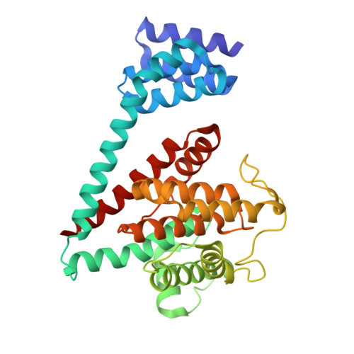
9YZ5 - PubMed Abstract:
FicD regulates Unfolded Protein Response (UPR) through reversible AMPylation and deAMPylation of BiP, an HSP70 chaperone and master regulator of the UPR. FicD activity is regulated by endoplasmic reticulum-stress, catalyzing BiP AMPylation under low stress conditions to hold inactive chaperone in reserve. In stressed cells, FicD deAMPylates BiP, acutely increasing its active pool to assist in protein folding. Variants in UPR machinery, including those in the FicD gene, are linked to hereditary diseases. Despite the known role of FicD in UPR, in-vivo regulation of its activity remains elusive, and identifying metabolites that alter FicD activity could prove useful pharmaceutically. We applied an unbiased high-throughput screening platform, known as Mass spectrometry Integrated with equilibrium Dialysis for the discovery of Allostery Systematically (MIDAS), to identify small molecule metabolites that might regulate FicD activity. MIDAS revealed interactions between FicD and two mevalonate pathway intermediates: geranyl-pyrophosphate and farnesyl-pyrophosphate. Biochemical characterization indicates that both potently inhibit FicD-mediated AMPylation and deAMPylation. The crystal structure of FicD bound to farnesyl-pyrophosphate demonstrates a competitive inhibition mechanism, with the pyrophosphate adopting the alpha and beta phosphate positions of adenosine triphosphate (ATP) and the hydrocarbon chain filling the nucleoside pocket. FicD variants previously appeared as biochemically indistinguishable, yet lead to different human pathologies. We demonstrate farnesyl-pyrophosphate inhibits FicD R374H and FicD R374C variants implicated in causing hereditary spastic paraplegia, but not the FicD R371S variant associated with neonatal diabetes. This study furthers our understanding of FicD inhibitors and distinguishes disease causing variants, providing insight into pharmacological targeting of UPR activity.
- Department of Molecular Biology, University of Texas Southwestern Medical Center, Dallas, TX 75390.
Organizational Affiliation: