Computational Design of a Highly Stable Dicopper Catechol Oxidase.
Eng, V.H., Narehood, S.M., Li, Y., Gascon, M., Hoffnagle, A.M., Shiau, A.A., Semonis, M., Green, M.T., Britt, R.D., Tezcan, F.A.(2026) J Am Chem Soc 148: 8361-8373
- PubMed: 41707222 Search on PubMed
- DOI: https://doi.org/10.1021/jacs.5c18979
- Primary Citation Related Structures:
9YTQ - PubMed Abstract:
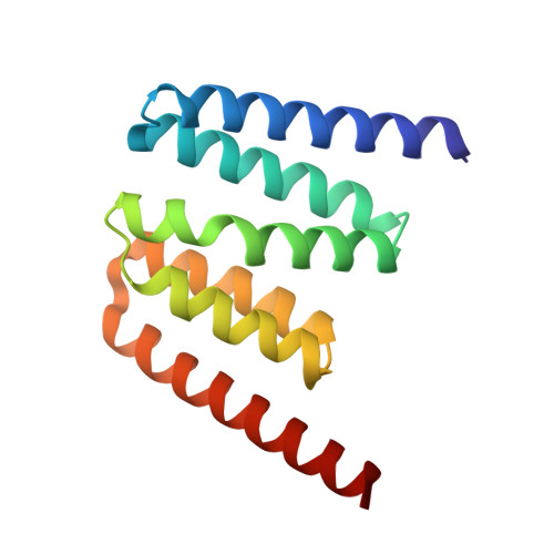
Type 3 (T3) Cu proteins play essential roles in binding and activating molecular oxygen (O 2 ) and are prevalent across all domains of life. Despite sharing the same coordination motif, T3 Cu proteins display divergent functions: hemocyanin transports O 2 , while tyrosinase catalyzes the hydroxylation of monophenols and the subsequent oxidation of diphenols and catechol oxidase oxidizes only diphenols. Here, we report the design and characterization of a di-Cu protein (Cu-HC4) inspired by the active sites of natural T3 Cu proteins to investigate the structural features that facilitate catalytic oxidase activity. Cu-HC4 is roughly 1/5th the size of the commercially available mushroom tyrosinase and shares only around 20% sequence identity with the T3 Cu protein templates. Notably, Cu-HC4 displays high thermostability and exhibits diphenol oxidation activity at ambient and elevated temperatures (≥60 °C). Cu-HC4 also initiates the formation of melanin polymers, mimicking melanin biosynthesis of natural tyrosinases. Mechanistic investigations demonstrate that Cu-HC4 utilizes both Cu centers cooperatively for diphenol oxidation and requires O 2 for catalysis like natural Cu oxidases but follows a distinct catalytic pathway compared to those enzymes. Cryo-EM characterization of a tetrameric form of HC4 reveals slight deviations in the relative positions of the active site His residues that may account for differences in reactivity between Cu-HC4 and natural T3 Cu enzymes.
- Department of Chemistry, University of California, San Diego, La Jolla, California 92093, United States.
Organizational Affiliation: