9YTF | pdb_00009ytf

TCR mimic antibody vAB-30 in complex with MAGE-A3 in HLA-A1


Experimental Data Snapshot

  • Method: ELECTRON MICROSCOPY
  • Resolution: 2.60 Å
  • Aggregation State: PARTICLE 
  • Reconstruction Method: SINGLE PARTICLE 

wwPDB Validation 3D Report Full Report

Validation slider image for 9YTF

This is version 1.0 of the entry. See complete history

Literature

Targeting peptide-MHC complexes with designed T cell receptors and antibodies.

Motmaen, A.Jude, K.M.Wang, N.Minervina, A.Feldman, D.Lichtenstein, M.A.Ebenezer, A.Correnti, C.Thomas, P.G.Garcia, K.C.Baker, D.Bradley, P.

(2025) bioRxiv 

  • DOI: https://doi.org/10.1101/2025.11.19.689381
  • Primary Citation Related Structures: 
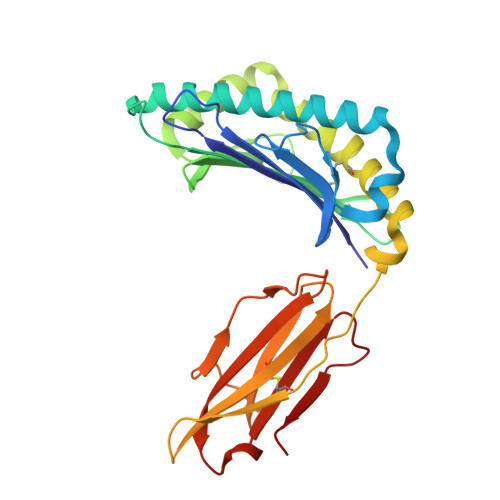
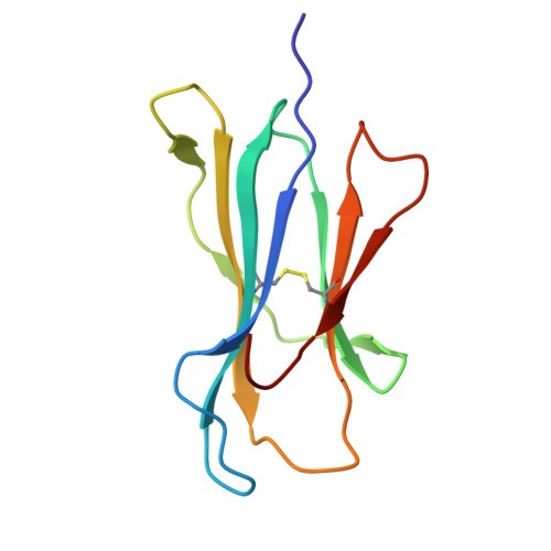
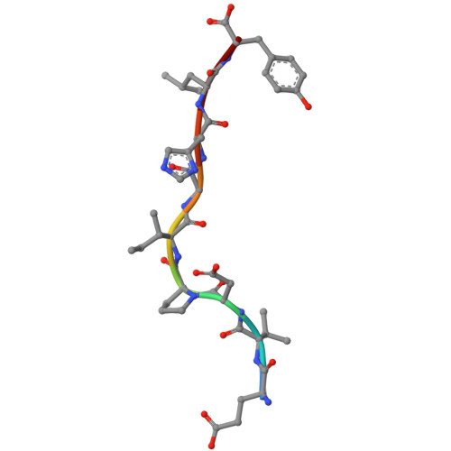
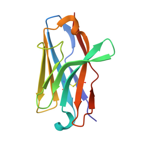
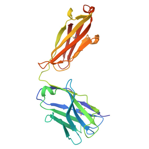
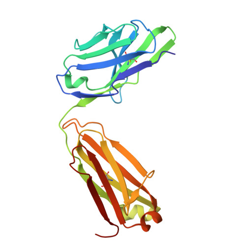
    9YTD, 9YTF

  • PubMed Abstract: 

    Class I major histocompatibility complexes (MHCs), expressed on the surface of all nucleated cells, present peptides derived from intracellular proteins for surveillance by T cells. The precise recognition of foreign or mutated peptide-MHC (pMHC) complexes by T cell receptors (TCRs) is central to immune defense against pathogens and tumors. Although patient-derived TCRs specific for cancer-associated antigens have been used to engineer tumor-targeting therapies, their reactivity toward self- or near-self antigens may be constrained by negative selection in the thymus. Here, we introduce a structure-based deep learning framework, ADAPT (Antigen-receptor Design Against Peptide-MHC Targets), for the design of TCRs and antibodies that bind to pMHC targets of interest. We evaluate the ADAPT pipeline by designing and characterizing TCRs and antibodies against a diverse panel of pMHCs. Cryogenic electron microscopy structures of two designed antibodies bound to their respective pMHC targets demonstrate atomic-level accuracy at the recognition interface, supporting the robustness of our structure-based approach. Computationally designed TCRs and antibodies targeting pMHC complexes could enable a broad range of therapeutic applications, from cancer immunotherapy to autoimmune disease treatment, and insights gained from TCR-pMHC design should advance predictive understanding of TCR specificity with implications for basic immunology and clinical diagnostics.


  • Organizational Affiliation
    • Institute for Protein Design, University of Washington, Seattle, WA, USA.

Macromolecule Content 

  • Total Structure Weight: 103.4 kDa 
  • Atom Count: 7,270 
  • Modeled Residue Count: 932 
  • Deposited Residue Count: 932 
  • Unique protein chains: 6

Macromolecules

Find similar proteins by:|  3D Structure
Entity ID: 1
MoleculeChains  Sequence LengthOrganismDetailsImage
MHC class I antigen275Homo sapiensMutation(s): 0 
Gene Names: HLA-A
UniProt
Find proteins for A0A1D3TZM3 (Homo sapiens)
Explore A0A1D3TZM3 
Go to UniProtKB:  A0A1D3TZM3
Entity Groups
Sequence Clusters30% Identity50% Identity70% Identity90% Identity95% Identity100% Identity
UniProt GroupA0A1D3TZM3
Sequence Annotations
Expand
Reference Sequence
Find similar proteins by:|  3D Structure
Entity ID: 2
MoleculeChains  Sequence LengthOrganismDetailsImage
Beta-2-microglobulin100Homo sapiensMutation(s): 0 
Gene Names: B2MCDABP0092HDCMA22P
UniProt & NIH Common Fund Data Resources
Find proteins for P61769 (Homo sapiens)
Explore P61769 
Go to UniProtKB:  P61769
PHAROS:  P61769
GTEx:  ENSG00000166710 
Entity Groups
Sequence Clusters30% Identity50% Identity70% Identity90% Identity95% Identity100% Identity
UniProt GroupP61769
Sequence Annotations
Expand
Reference Sequence
Find similar proteins by:  Sequence   |   3D Structure  
Entity ID: 3
MoleculeChains  Sequence LengthOrganismDetailsImage
Melanoma-associated antigen 39Homo sapiensMutation(s): 0 
Gene Names: MAGEA3MAGE3
UniProt & NIH Common Fund Data Resources
Find proteins for P43357 (Homo sapiens)
Explore P43357 
Go to UniProtKB:  P43357
PHAROS:  P43357
GTEx:  ENSG00000221867 
Entity Groups
UniProt GroupP43357
Sequence Annotations
Expand
Reference Sequence
Find similar proteins by:|  3D Structure
Entity ID: 4
MoleculeChains  Sequence LengthOrganismDetailsImage
AD01-VHH115Homo sapiensMutation(s): 0 
Find similar proteins by:|  3D Structure
Entity ID: 5
MoleculeChains  Sequence LengthOrganismDetailsImage
vAB30 light chainE [auth F]218synthetic constructMutation(s): 0 
Find similar proteins by:|  3D Structure
Entity ID: 6
MoleculeChains  Sequence LengthOrganismDetailsImage
vAB30 heavy chainF [auth G]215synthetic constructMutation(s): 0 

Experimental Data & Validation

Experimental Data

  • Method: ELECTRON MICROSCOPY
  • Resolution: 2.60 Å
  • Aggregation State: PARTICLE 
  • Reconstruction Method: SINGLE PARTICLE 
EM Software:
TaskSoftware PackageVersion
MODEL REFINEMENTPHENIX1.20.1_4487:
RECONSTRUCTIONcryoSPARC

Structure Validation

View Full Validation Report


Entry History 

& Funding Information

Deposition Data


Funding OrganizationLocationGrant Number
Cancer Research UKUnited KingdomCGCATF-2023/100006

Revision History  (Full details and data files)

  • Version 1.0: 2025-12-17
    Type: Initial release