Download Mendeley
Vaccination elicits HIV broadly neutralizing antibodies in primates.
Steichen, J.M., Madden, P.J., Flynn, C.T., Phulera, S., Shil, M., Kalyuzhniy, O., Liguori, A., Kifude, C., Sewall, L.M., Cottrell, C.A., Ma, K.M., Baboo, S., Diedrich, J.K., McKenney, K., deCamp, A.C., Carnathan, D.G., Phung, I., Ramezani-Rad, P., Marina-Zarate, E., Freeman, B., Xie, Z., Lee, J.H., Sincomb, T., Phelps, N., Lu, D., Goodwin, D., Tingle, R., Adachi, Y., Alavi, N., Tran, J., Tran, A.S., Nascimento, A., Sovie, C., Bader, D.L.V., Voic, H., Zhou, X., Pixton, G., Walsh, A., Melo, M.B., Schiffner, T., Batista, F.D., Burton, D.R., Irvine, D.J., Paulson, J.C., Yates 3rd, J.R., Ozorowski, G., Ward, A.B., Silvestri, G., Crotty, S., Schief, W.R.(2026) Nature 656: 723-733
- PubMed: 42380658 Search on PubMedSearch on PubMed Central
- DOI: https://doi.org/10.1038/s41586-026-10837-5
- Primary Citation Related Structures:
9YMJ, 9YMK, 9YML - PubMed Abstract:
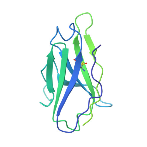
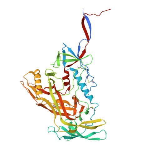
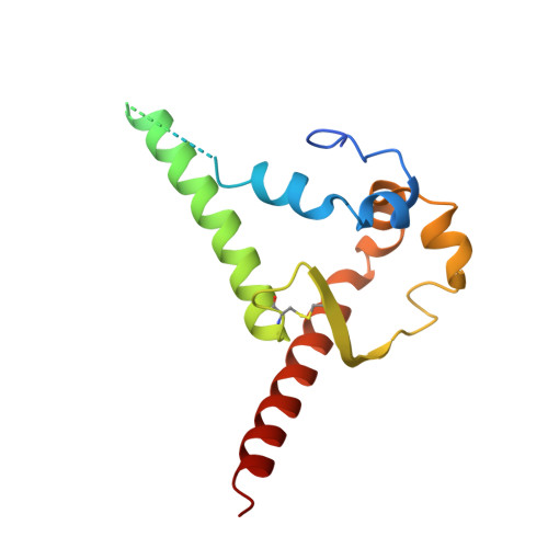
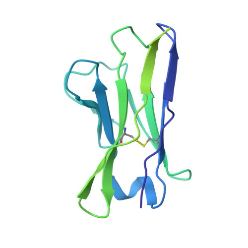
The high antigenic diversity of HIV has been a major obstacle to development of a broadly protective vaccine. Nevertheless, protective HIV broadly neutralizing antibodies (bnAbs) exist and have been proposed as templates for vaccine development 1-6 . Germline targeting is a conceptually radical vaccine design approach to elicit bnAbs, aiming to prime rare bnAb-precursor B cells possessing predetermined human genetic and structural features shared with template bnAbs, and then guide B cell affinity maturation to potent bnAb evolution with heterologous boosters 7-11 . Although the approach has shown promise in clinical 12-17 and preclinical 18-34 studies, it faces many immunological challenges and, to date, has not succeeded in generating bnAbs in humans or non-transgenic animals. Here we report testing of an adjuvanted protein germline-targeting vaccine in outbred non-human primates. The vaccine generated bnAb-class memory B cells and sera capable of neutralizing diverse HIV clinical isolates. bnAb lineages were generated in at least 50% of animals, achieving up to 67% neutralization breadth compared with the reference bnAb. Vaccine-induced bnAbs exhibited precise structural mimicry of human bnAb interactions with HIV envelope (Env), matching the germline-targeting predictions. Furthermore, serum bnAb activity developed in 44% of animals and in one instance reached titres expected to confer protection against diverse HIV isolates. These results demonstrate proof of principle that germline-targeting vaccines can reproducibly elicit prespecified classes of bnAbs to prespecified epitopes under endogenous conditions, supporting further optimization of this approach for HIV vaccine development.
- Department of Immunology and Microbiology, The Scripps Research Institute, La Jolla, CA, USA.
Organizational Affiliation: