Find PDB structures and Computed Structure Models (CSM) by combining queries from tools in this suite: Attribute Search, Sequence Similarity, Sequence Motif, 3D Similarity, and 3D Motif with 'AND' logic.
Funding Organization(s): National Institutes of Health/National Institute of General Medical Sciences (NIH/NIGMS), National Institutes of Health/National Center for Complementary and Integrative Health (NIH/NCCIH)
Primary Citation Related Structures: 9YH3, 9YH4, 9YH5
PubMed Abstract:
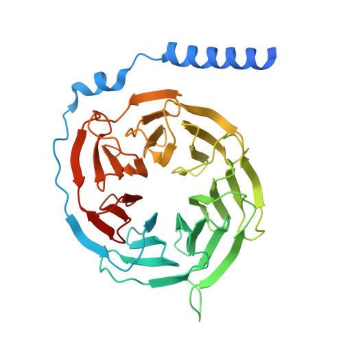
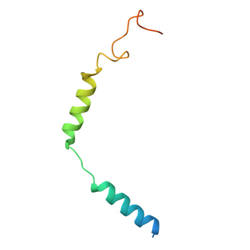
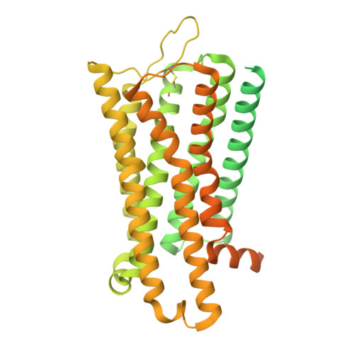
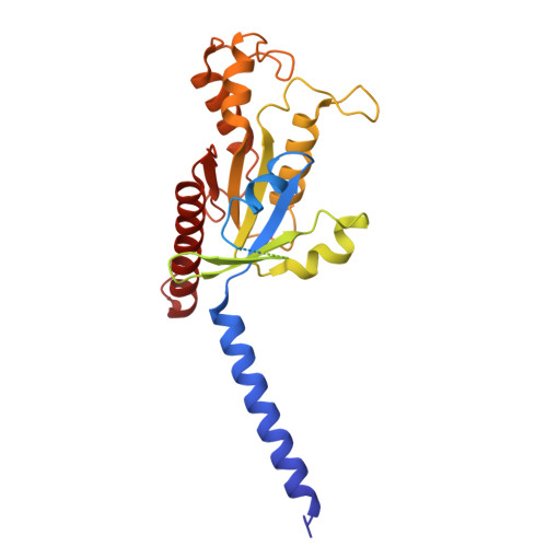
The mu-opioid receptor (MOR) is a major target for pain treatment, but its effectiveness is limited by dangerous side effects. However, the systemic administration of opioids makes it difficult both to decipher their underlying mechanisms of action and to limit drug action to specific receptor subpopulations to isolate therapeutic effects from adverse effects. Here, we develop a reversibly photoswitchable morphinan agonist termed "azo-morphine-3" (AM-3) that interconverts from low to high efficacy in response to light to enable optical control of MOR signaling. Structural analysis of the low ("trans") and high ("cis") efficacy states of AM-3 bound to the MOR reveals distinct binding modes, with each state driving unique ligand, receptor, and G protein dynamics. In mice, AM-3 drives reversible and repeatable optical control of antinociception across pain models with a reduced side effect profile owing to its limited brain accumulation and its ability to be locally activated.
Organizational Affiliation:
Department of Biochemistry & Biophysics, Weill Cornell Medicine, New York, NY 10065, USA.
Department of Chemistry, University of Pennsylvania, Philadelphia, PA 19104, USA.
The Bridge Institute, Michelson Center for Convergent Biosciences, University of Southern California, Los Angeles, CA 90089, USA; Department of Molecular and Cellular Biosciences, University of Southern California, Los Angeles, CA 90089, USA.
The Bridge Institute, Michelson Center for Convergent Biosciences, University of Southern California, Los Angeles, CA 90089, USA; Department of Quantitative and Computational Biology, University of Southern California, Los Angeles, CA 90089, USA.
Department of Chemistry, New York University, New York, NY 10003, USA; Department of Fundamental Chemistry, Institute of Chemistry, University of São Paulo, Avenida Professor Lineu Prestes, 748, 05508-000 São Paulo, Brazil.
The Bridge Institute, Michelson Center for Convergent Biosciences, University of Southern California, Los Angeles, CA 90089, USA.
Department of Molecular and Cellular Biosciences, University of Southern California, Los Angeles, CA 90089, USA; Department of Quantitative and Computational Biology, University of Southern California, Los Angeles, CA 90089, USA.
The Bridge Institute, Michelson Center for Convergent Biosciences, University of Southern California, Los Angeles, CA 90089, USA; Keck School of Medicine, University of Southern California, Los Angeles, CA, USA.
The Bridge Institute, Michelson Center for Convergent Biosciences, University of Southern California, Los Angeles, CA 90089, USA; Department of Chemistry, University of Southern California, Los Angeles, CA 90089, USA.
Department of Pharmacology, Weill Cornell Medicine, New York, NY 10065, USA.
Molecular Biophysics and Integrated Bio-Imaging Division, Lawrence Berkeley National Laboratory, Berkeley, CA 94720-8142, USA.
The Bridge Institute, Michelson Center for Convergent Biosciences, University of Southern California, Los Angeles, CA 90089, USA; Department of Quantitative and Computational Biology, University of Southern California, Los Angeles, CA 90089, USA; Department of Chemistry, University of Southern California, Los Angeles, CA 90089, USA. Electronic address: katritch@usc.edu.
The Bridge Institute, Michelson Center for Convergent Biosciences, University of Southern California, Los Angeles, CA 90089, USA; Department of Molecular and Cellular Biosciences, University of Southern California, Los Angeles, CA 90089, USA; Department of Chemistry, University of Southern California, Los Angeles, CA 90089, USA. Electronic address: gati@usc.edu.
Department of Chemistry, University of Pennsylvania, Philadelphia, PA 19104, USA; Department of Systems Pharmacology and Translational Therapeutics, Perelman School of Medicine, University of Pennsylvania, Philadelphia, PA 19104, USA. Electronic address: dtrauner@upenn.edu.
Department of Biochemistry & Biophysics, Weill Cornell Medicine, New York, NY 10065, USA; Department of Psychiatry, Weill Cornell Medicine, New York, NY 10065, USA. Electronic address: jtl2003@med.cornell.edu.

 Download Mendeley