Assembly and glycosylation of Helicobacter pylori sheathed flagella.
Kumar, R., Tachiyama, S., Yu, H., Heydari, S., Guo, J., Botting, J.M., Guo, W., Hoover, T.R., Liu, J.(2026) PNAS Nexus 5: pgag011-pgag011
- PubMed: 41659214 Search on PubMedSearch on PubMed Central
- DOI: https://doi.org/10.1093/pnasnexus/pgag011
- Primary Citation Related Structures:
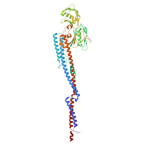
9YGU, 9YH1 - PubMed Abstract:
The bacterial flagellum is a complex nanomachine essential for motility, colonization, and invasion in diverse species. Helicobacter pylori has evolved elaborate sheathed flagella that enable migration through the highly viscous gastric mucus layer to reach its colonization niche on the gastric epithelium, yet the molecular basis for these unique adaptations has remained elusive. Here, we use in situ single-particle cryo-electron microscopy to determine near-atomic structures of the flagellar filament within the membranous sheath of H. pylori . The major flagellin FlaA constitutes the bulk of the filament, whereas the minor flagellin FlaB contributes critically to the hook-proximal region. Both FlaA and FlaB form a conserved core surrounded by variable surface-exposed domains. Our structures further reveal that pseudaminic acid glycans decorate these domains, where they mediate inter- and intra-subunit contacts that stabilize the filament and confer a negatively charged surface. Together, these findings support a model in which the filament rotates independently of the membranous sheath to drive H. pylori motility and provide a molecular framework for understanding how the sheathed flagellum enables colonization and persistence within the gastric niche.
- Department of Microbial Pathogenesis, Yale School of Medicine, New Haven, CT 06536, USA.
Organizational Affiliation: