Targeting PTPN22 at Nonorthosteric Binding SitesA Fragment Approach.
Di Lello, P., Wells, M.M., Davis, B., Daniels, Z., Garner, T.P., Gazzard, L., Harris, R., Hubbard, R.E., Landry, M.L., Martin, B., Morgan, J.L.W., Patapoff, A., Simmonite, H., Skelton, N., Ultsch, M., Walters, B.T., Wu, P., Dimitrova, Y.N., Huard, K.(2026) ACS Omega 11: 3465-3480
- PubMed: 41585704 Search on PubMedSearch on PubMed Central
- DOI: https://doi.org/10.1021/acsomega.5c11028
- Primary Citation Related Structures:
9YDM, 9YG0, 9YG1, 9YG2, 9YG3 - PubMed Abstract:
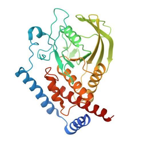
Nonreceptor protein tyrosine phosphatase 22 (PTPN22) is a known negative regulator of T cell receptor signaling. PTPN22's pro-autoimmune variant (C1858T) was found to have a risk preventive association with multiple types of cancer, to contribute to improved overall survival in patients treated with the anti-PD-L1 atezolizumab, and to enhance tumor immunity in mice. Modulating the activity of phosphatases has been historically challenging due to the polar and conserved nature of the orthosteric sites across the protein family. In this work, we outline a strategy for discovering and characterizing nonorthosteric ligands of the PTPN22 phosphatase domain. We opted for a fragment screen to identify ligands of PTPN22 and utilized a multidisciplinary approach to characterize them. This included the integration of experimental data-driven molecular dynamics when cocrystallization of fragments with PTPN22 was unsuccessful. With this approach, we identified and advanced fragments that bind PTPN22 at two novel nonorthosteric sites. Due to the shared tertiary structure of the phosphatase domain, we believe this hit finding effort, combined with knowledge about the allosteric circuitry of phosphatases, can provide synergistic value.
- Department of Structural Biology, Genentech, Inc., 1 DNA Way, South San Francisco, California 94080, United States.
Organizational Affiliation: