9YCP | pdb_00009ycp

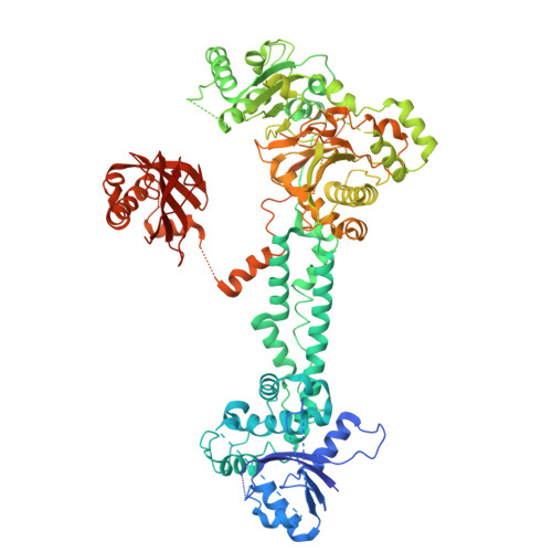
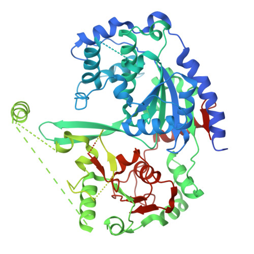
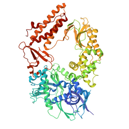
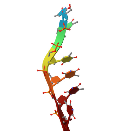
HSV Helicase-primase complex bound to IM-250


Experimental Data Snapshot

  • Method: ELECTRON MICROSCOPY
  • Resolution: 2.90 Å
  • Aggregation State: PARTICLE 
  • Reconstruction Method: SINGLE PARTICLE 

wwPDB Validation 3D Report Full Report

Validation slider image for 9YCP

This is version 1.1 of the entry. See complete history

Literature

Mechanisms of HSV-1 helicase-primase inhibition and replication fork complex assembly.

Yu, Z.Sathyanarayana, P.Liu, C.Tan, J.M.J.Yang, P.Das, B.Hu, S.Fan, X.Ji, C.Weller, S.K.Shekhar, M.Coen, D.M.Kranzusch, P.J.Loparo, J.J.Abraham, J.

(2026) Cell 189: 478-494.e18

  • DOI: https://doi.org/10.1016/j.cell.2025.11.041
  • Primary Citation Related Structures: 
    9YC9, 9YCP, 9YCT, 9YCV

  • PubMed Abstract: 

    Herpesviruses are widespread double-stranded DNA viruses that establish lifelong latency and cause various diseases. Although DNA-polymerase-targeting antivirals are effective, increasing drug resistance underscores the need for alternatives. Helicase-primase inhibitors (HPIs) are promising antivirals, but their mechanisms of action are poorly defined. Furthermore, how the helicase-primase (H/P) complex and DNA polymerase coordinate genome replication is not well understood for herpesviruses. Here, we report cryo-electron microscopy (cryo-EM) structures of the herpes simplex virus 1 H/P complex bound to HPIs, showing that these lock the H/P complex in an inactive state. Single-molecule assays reveal that HPIs cause H/P complexes to pause in unwinding activity on DNA. The structure of an HPI-bound replication fork complex, comprising the H/P complex (UL5, UL52, and UL8) and the polymerase holoenzyme (UL30 and UL42), reveals a previously uncharacterized interface bridging these complexes. These findings provide a structural framework for understanding herpesvirus replisome assembly and advancing inhibitor development.


  • Organizational Affiliation
    • Department of Microbiology, Blavatnik Institute, Harvard Medical School, Boston, MA, USA.

Macromolecule Content 

  • Total Structure Weight: 295.67 kDa 
  • Atom Count: 15,394 
  • Modeled Residue Count: 1,977 
  • Deposited Residue Count: 2,696 
  • Unique protein chains: 3
  • Unique nucleic acid chains: 1

Macromolecules


Find similar proteins by:|  3D Structure
Entity ID: 2
MoleculeChains  Sequence LengthOrganismDetailsImage
UL52B [auth C]1,058Human alphaherpesvirus 1 strain R-15Mutation(s): 0 
Gene Names: UL52
UniProt
Find proteins for A0A5J6DWG4 (Human herpesvirus 1)
Explore A0A5J6DWG4 
Go to UniProtKB:  A0A5J6DWG4
Entity Groups
Sequence Clusters30% Identity50% Identity70% Identity90% Identity95% Identity100% Identity
UniProt GroupA0A5J6DWG4
Sequence Annotations
Expand
Reference Sequence
Find similar proteins by:|  3D Structure
Entity ID: 3
MoleculeChains  Sequence LengthOrganismDetailsImage
UL5C [auth A]882Human alphaherpesvirus 1 strain R-15Mutation(s): 0 
Gene Names: UL5
UniProt
Find proteins for A0A5J6DVR7 (Human herpesvirus 1)
Explore A0A5J6DVR7 
Go to UniProtKB:  A0A5J6DVR7
Entity Groups
Sequence Clusters30% Identity50% Identity70% Identity90% Identity95% Identity100% Identity
UniProt GroupA0A5J6DVR7
Sequence Annotations
Expand
Reference Sequence
Find similar proteins by:|  3D Structure
Entity ID: 4
MoleculeChains  Sequence LengthOrganismDetailsImage
UL8D [auth B]750Human alphaherpesvirus 1 strain R-15Mutation(s): 0 
Gene Names: UL8
UniProt
Find proteins for P10192 (Human herpesvirus 1 (strain 17))
Explore P10192 
Go to UniProtKB:  P10192
Entity Groups
Sequence Clusters30% Identity50% Identity70% Identity90% Identity95% Identity100% Identity
UniProt GroupP10192
Sequence Annotations
Expand
Reference Sequence
Find similar nucleic acids by:  Sequence
Entity ID: 1
MoleculeChains LengthOrganismImage
DNA (5'-D(*TP*TP*TP*TP*TP*T)-3')A [auth D]6Human alphaherpesvirus 1 strain R-15
Sequence Annotations
Expand
Reference Sequence

Small Molecules

Ligands 2 Unique
IDChains Name / Formula / InChI Key2D Diagram3D Interactions
A1CHB
(Subject of Investigation/LOI)

Query on A1CHB


Download:Ideal Coordinates CCD File
F [auth C]N-{5-[(R)-amino(hydroxy)(methyl)-lambda~4~-sulfanyl]-4-methyl-1,3-thiazol-2-yl}-2-(2',5'-difluoro[1,1'-biphenyl]-4-yl)-N-methylacetamide
C20 H21 F2 N3 O2 S2
XMZIBPDSCWYNDP-UHFFFAOYSA-N
ZN

Query on ZN


Download:Ideal Coordinates CCD File
E [auth C]ZINC ION
Zn
PTFCDOFLOPIGGS-UHFFFAOYSA-N

Experimental Data & Validation

Experimental Data

  • Method: ELECTRON MICROSCOPY
  • Resolution: 2.90 Å
  • Aggregation State: PARTICLE 
  • Reconstruction Method: SINGLE PARTICLE 
EM Software:
TaskSoftware PackageVersion
RECONSTRUCTIONcryoSPARC

Structure Validation

View Full Validation Report


Entry History 

& Funding Information

Deposition Data


Funding OrganizationLocationGrant Number
Not funded--

Revision History  (Full details and data files)

  • Version 1.0: 2025-12-31
    Type: Initial release
  • Version 1.1: 2026-07-15
    Changes: Data collection, Database references