Oligomeric defects in soybean serine hydroxymethyltransferase 8: tetramer destabilization by A149T and other variants associated with soybean cyst nematode resistance.
Samarakoon, V., Buckley, D.P., Owuocha, L.F., Durie, C.L., Mitchum, M.G., Beamer, L.J.(2026) FEBS J
- PubMed: 41914311 Search on PubMed
- DOI: https://doi.org/10.1111/febs.70528
- Primary Citation Related Structures:
10NM, 9Y6B, 9Y6D, 9Y7G, 9YBZ - PubMed Abstract:
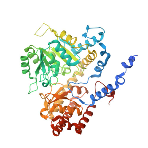
Serine hydroxymethyltransferase (SHMT) is a conserved enzyme in folate-mediated one-carbon metabolism, where it contributes to nucleotide biosynthesis, methylation capacity, and cellular stress responses. Amino acid polymorphisms of soybean SHMT8 are known to affect the resistance of soybean to its primary pathogen, the soybean cyst nematode (SCN). A set of SHMT8 variants from ethyl methanesulfonate (EMS)-mutagenized soybean populations has been identified with varying resistance phenotypes, but their biochemical consequences remain poorly understood. Here, we use biochemical and structural studies to assess the impacts of the A149T variant on soybean SHMT8. Despite the conservative nature of the substitution, A149T reduces folate binding, pyridoxal-5'-phosphate-dependent catalysis, and thermal stability. High-resolution crystal structures (1.9-2.3 Å resolution) reveal only very minor structural changes. However, while the usual tetrameric assembly of the enzyme is retained at the high protein concentration in crystals, multiple other methods including a 2.9 Å cryo-electron microscopy (cryo-EM) structure show that the A149T variant is predominantly a dimer. Significant structural changes in the dimer are consistent with the observed biochemical impacts of the variant and help explain the well-known reduction in activity associated with dimerization of SHMT in other systems. We also find destabilization of the tetrameric assembly in other SHMT8 variants associated with changes in SCN resistance, suggesting that weakened oligomerization may be a common consequence of such mutations. Together, these results highlight quaternary structure as a critical determinant of SHMT8 activity and stability and suggest a potential mechanistic link between enzyme biochemistry and soybean defense.
- Department of Chemistry, University of Missouri, Columbia, MO, USA.
Organizational Affiliation: