9Y68 | pdb_00009y68

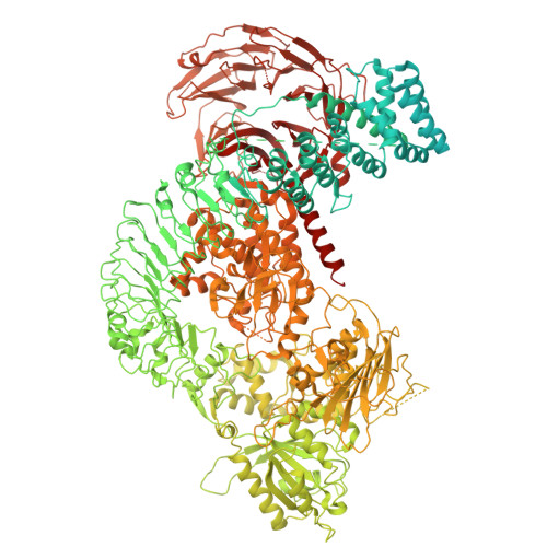
Full length LRRK2 (G2019S) after symmetry expansion


Experimental Data Snapshot

  • Method: ELECTRON MICROSCOPY
  • Resolution: 3.25 Å
  • Aggregation State: PARTICLE 
  • Reconstruction Method: SINGLE PARTICLE 

wwPDB Validation 3D Report Full Report

Validation slider image for 9Y68

This is version 1.1 of the entry. See complete history

Literature

The structural basis for LRRK2's activation and autoinhibition.

Villagran Suarez, A.Hatch, K.S.Bodrug, T.Gai, W.Surridge, K.J.Moussikhina, E.Nguyen, K.H.V.Sanz-Murillo, M.Callahan, R.Xiong, E.Ramos, D.Zhu, L.Dederer, V.Mathea, S.Iwasa, J.Knapp, S.Shokat, K.M.Reck-Peterson, S.L.Leschziner, A.E.

(2026) Cell 

  • DOI: https://doi.org/10.1016/j.cell.2026.07.027
  • Primary Citation Related Structures: 
    9OM2, 9OXH, 9OXI, 9OYA, 9Y67, 9Y68, 9YQK

  • PubMed Abstract: 

    Mutations in leucine-rich repeat kinase 2 (LRRK2) are the second most common cause of autosomal-dominant Parkinson's disease (PD), and increased LRRK2 kinase activity is also observed in idiopathic PD, making LRRK2 a major actionable therapeutic target. LRRK2 is a 286-kDa multidomain enzyme containing a Ras-like GTPase (ROC) and a kinase domain. Using cryo-electron microscopy (cryo-EM), biochemical reconstitution, and cell-based assays, we show that the ROC GTPase governs switching between autoinhibited and active states: GTP binding promotes activation, whereas GDP binding enforces autoinhibition. Two common PD-linked mutations, G2019S and R1441C/G/H, activate LRRK2 through distinct structural mechanisms, revealing genotype-specific routes to dysregulation. These findings provide a unified framework for understanding LRRK2 regulation with broad therapeutic implications. Stabilizing the guanosine diphosphate (GDP)-bound state may inhibit LRRK2 by maintaining autoinhibition, whereas promoting the GTP-bound state could be advantageous in specific cellular contexts, such as the lung, where increased LRRK2 kinase activity may play protective or regulatory roles.


  • Organizational Affiliation
    • Department of Cellular and Molecular Medicine, University of California, San Diego, La Jolla, CA 92093, USA.

Macromolecule Content 

  • Total Structure Weight: 286.84 kDa 
  • Atom Count: 13,046 
  • Modeled Residue Count: 1,700 
  • Deposited Residue Count: 2,527 
  • Unique protein chains: 1

Macromolecules

Find similar proteins by:|  3D Structure
Entity ID: 1
MoleculeChains  Sequence LengthOrganismDetailsImage
Leucine-rich repeat serine/threonine-protein kinase 22,527Homo sapiensMutation(s): 1 
Gene Names: LRRK2PARK8
EC: 2.7.11.1 (PDB Primary Data), 3.6.5 (PDB Primary Data)
UniProt & NIH Common Fund Data Resources
Find proteins for Q5S007 (Homo sapiens)
Explore Q5S007 
Go to UniProtKB:  Q5S007
GTEx:  ENSG00000188906 
Entity Groups
Sequence Clusters30% Identity50% Identity70% Identity90% Identity95% Identity100% Identity
UniProt GroupQ5S007
Sequence Annotations
Expand
Reference Sequence

Small Molecules

Ligands 1 Unique
IDChains Name / Formula / InChI Key2D Diagram3D Interactions
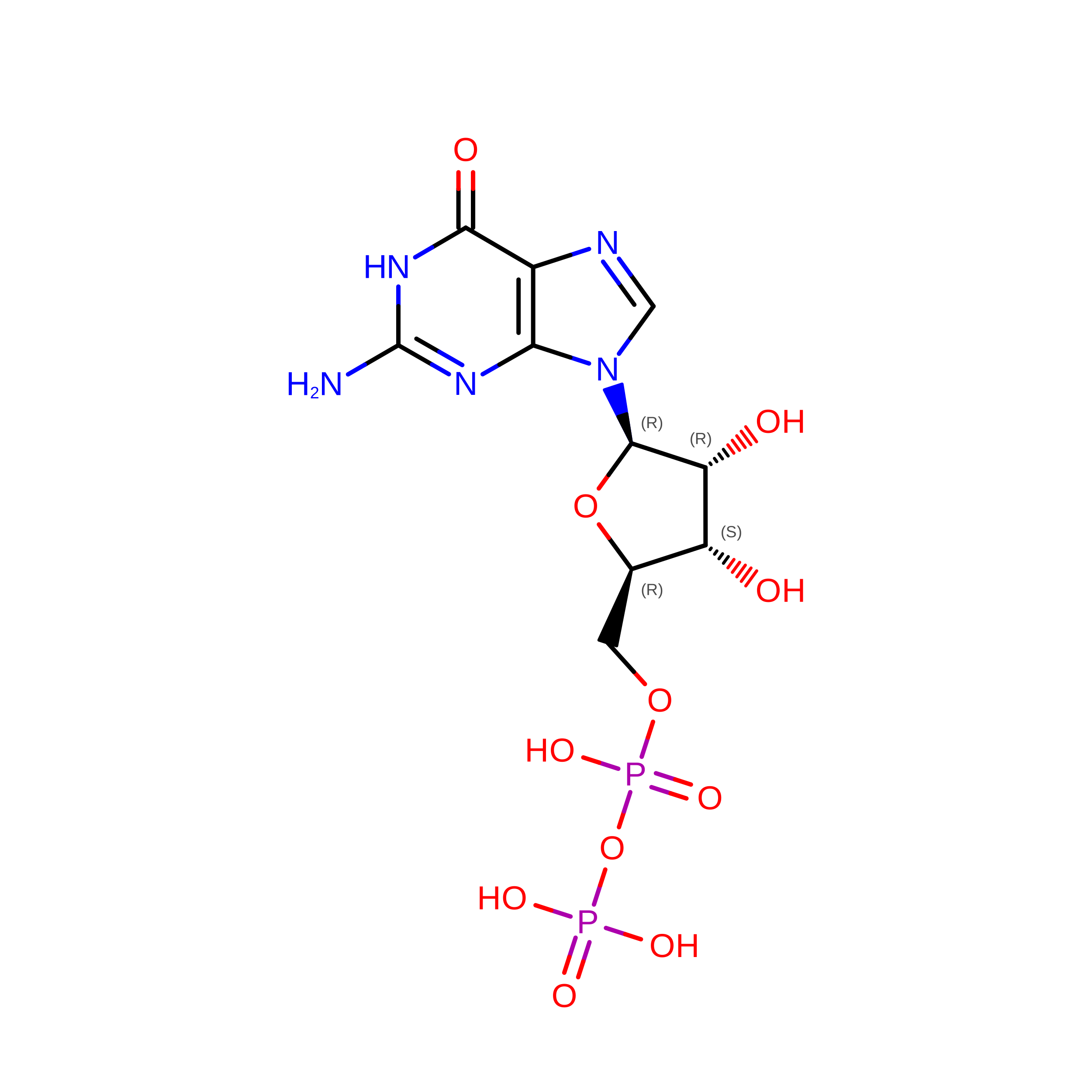
GDP
(Subject of Investigation/LOI)

Query on GDP


Download:Ideal Coordinates CCD File
B [auth A]GUANOSINE-5'-DIPHOSPHATE
C10 H15 N5 O11 P2
QGWNDRXFNXRZMB-UUOKFMHZSA-N

Experimental Data & Validation

Experimental Data

  • Method: ELECTRON MICROSCOPY
  • Resolution: 3.25 Å
  • Aggregation State: PARTICLE 
  • Reconstruction Method: SINGLE PARTICLE 
EM Software:
TaskSoftware PackageVersion
MODEL REFINEMENTPHENIX1.20.1_4487
RECONSTRUCTIONcryoSPARC

Structure Validation

View Full Validation Report


Entry History 

& Funding Information

Deposition Data


Funding OrganizationLocationGrant Number
Michael J. Fox FoundationUnited StatesASAP-000519

Revision History  (Full details and data files)

  • Version 1.0: 2026-08-12
    Type: Initial release
  • Version 1.1: 2026-08-19
    Changes: Data collection, Database references