9Y2Z | pdb_00009y2z

Icosahedral symmetric structure of an expansion intermediate of Turnip Crinkle Virus (Asymmetric Trimer Unit)


Experimental Data Snapshot

  • Method: ELECTRON MICROSCOPY
  • Resolution: 2.87 Å
  • Aggregation State: PARTICLE 
  • Reconstruction Method: SINGLE PARTICLE 

Starting Model: experimental
View more details

wwPDB Validation   3D Report Full Report


This is version 1.1 of the entry. See complete history


Literature

Asymmetric isopeptide bond steers directional genomic RNA egress from icosahedral virus.

Braet, S.M.Venkatakrishnan, V.Ramesh, R.Clawson, M.A.Laremore, T.N.Wong, S.M.Anand, G.S.

(2025) Sci Adv 11: eady4104-eady4104

  • DOI: https://doi.org/10.1126/sciadv.ady4104
  • Primary Citation of Related Structures:  
    9Y2Z, 9Y31

  • PubMed Abstract: 

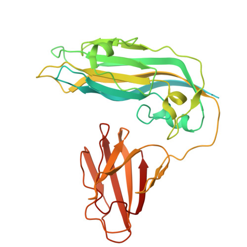
    Icosahedral RNA viruses rely upon essential asymmetries for directional genome egress into the host cell. How these asymmetric egress points are built into the quaternary assembly of the virion is unknown. Here, we capture the structure and dynamics of a partially expanded virus disassembly intermediate, poised to release its spring-loaded genomic RNA. The structure shows highly localized density of RNA underneath surface fivefold axes on one-half of the viral particle. This polarity in RNA distribution is associated with a unique interchain isopeptide bond (glutamic acid-lysine), which flanks conserved high-affinity RNA binding sites. This singular isopeptide bond at the asymmetric egress site confers an essential "loaded-die" feature that is critical for steering genomic RNA egress into the host cell for virus propagation.


  • Organizational Affiliation
    • Department of Chemistry, Penn State University, University Park, PA 16802, USA.

Macromolecules
Find similar proteins by:  (by identity cutoff)  |  3D Structure
Entity ID: 1
MoleculeChains Sequence LengthOrganismDetailsImage
Capsid protein
A, B, C
351Turnip crinkle virusMutation(s): 0 
UniProt
Find proteins for P06663 (Turnip crinkle virus)
Explore P06663 
Go to UniProtKB:  P06663
Entity Groups  
Sequence Clusters30% Identity50% Identity70% Identity90% Identity95% Identity100% Identity
UniProt GroupP06663
Sequence Annotations
Expand
  • Reference Sequence
Experimental Data & Validation

Experimental Data

  • Method: ELECTRON MICROSCOPY
  • Resolution: 2.87 Å
  • Aggregation State: PARTICLE 
  • Reconstruction Method: SINGLE PARTICLE 
EM Software:
TaskSoftware PackageVersion
RECONSTRUCTIONcryoSPARC4.6
MODEL REFINEMENTPHENIX

Structure Validation

View Full Validation Report


Entry History & Funding Information

Deposition Data


Funding OrganizationLocationGrant Number
National Institutes of Health/National Institute of General Medical Sciences (NIH/NIGMS)United StatesR35 GM158169-01

Revision History  (Full details and data files)

  • Version 1.0: 2025-11-12
    Type: Initial release
  • Version 1.1: 2025-12-24
    Changes: Data collection, Database references