9Y0N | pdb_00009y0n

Crystal structure of Escherichia coli DsbA G149K mutant


Experimental Data Snapshot

  • Method: X-RAY DIFFRACTION
  • Resolution: 2.00 Å
  • R-Value Free: 
    0.243 (Depositor), 0.241 (DCC) 
  • R-Value Work: 
    0.203 (Depositor), 0.202 (DCC) 
  • R-Value Observed: 
    0.205 (Depositor) 

Starting Model: experimental
View more details

wwPDB Validation 3D Report Full Report

Validation slider image for 9Y0N

This is version 1.1 of the entry. See complete history

Literature

A universal cis-proline lock defines catalysis in thioredoxin-fold enzymes.

Cunliffe, T.Wang, G.Penning, S.Subedi, P.Totsika, M.Paxman, J.J.Heras, B.

(2026) Commun Biol 

  • DOI: https://doi.org/10.1038/s42003-026-10010-8
  • Primary Citation Related Structures: 
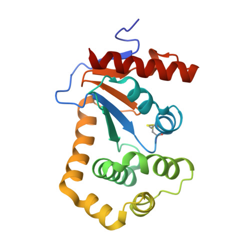
    9Y0M, 9Y0N, 9Y0O, 9Y0P, 9Y0Q

  • PubMed Abstract: 

    Thioredoxin-fold oxidoreductases drive oxidative protein folding and redox homeostasis across all domains of life. They catalyse thiol-disulfide exchange in diverse substrates, yet how they reconcile catalytic precision with substrate diversity remains unclear. Here we show, using high-resolution structures and functional analyses of the Escherichia coli oxidoreductase DsbA, that a conserved cis-proline loop adjacent to the catalytic Cys-Pro-His-Cys motif serves as a universal catalytic lock. The loop positions the substrate cysteine in a right-handed disulfide geometry optimal for exchange, while surrounding surfaces accommodate sequence variation. Substitution of the cis-proline abolishes turnover, whereas mutation of the preceding glycine preserves geometry but reduces efficiency. Comparative structural analyses demonstrate that this cis-proline-dependent hydrogen-bonding scaffold is conserved across thioredoxins, protein disulfide isomerases, peroxiredoxins and bacterial Dsb proteins. This conserved mechanism explains how catalytic fidelity is maintained while enabling substrate versatility and provides a foundation for enzyme engineering and therapeutic development.


  • Organizational Affiliation
    • Department of Biochemistry and Chemistry, School of Agriculture, Biomedicine and Environment, La Trobe Institute for Molecular Science, La Trobe University, Melbourne, VIC, Australia.

Macromolecule Content 

  • Total Structure Weight: 43.18 kDa 
  • Atom Count: 3,093 
  • Modeled Residue Count: 374 
  • Deposited Residue Count: 380 
  • Unique protein chains: 1

Macromolecules

Find similar proteins by:|  3D Structure
Entity ID: 1
MoleculeChains  Sequence LengthOrganismDetailsImage
Thiol:disulfide interchange protein DsbA
A, B
190Escherichia coli K-12Mutation(s): 1 
Gene Names: dsbAdsfppfAb3860JW3832
UniProt
Find proteins for P0AEG4 (Escherichia coli (strain K12))
Explore P0AEG4 
Go to UniProtKB:  P0AEG4
Entity Groups
Sequence Clusters30% Identity50% Identity70% Identity90% Identity95% Identity100% Identity
UniProt GroupP0AEG4
Sequence Annotations
Expand
Reference Sequence

Experimental Data & Validation

Experimental Data

  • Method: X-RAY DIFFRACTION
  • Resolution: 2.00 Å
  • R-Value Free:  0.243 (Depositor), 0.241 (DCC) 
  • R-Value Work:  0.203 (Depositor), 0.202 (DCC) 
  • R-Value Observed: 0.205 (Depositor) 
Space Group: P 21 21 21
Unit Cell:
Length ( Å )Angle ( ˚ )
a = 68.887α = 90
b = 79.301β = 90
c = 82.428γ = 90
Software Package:
Software NamePurpose
PHENIXrefinement
Aimlessdata scaling
PHASERphasing
PDB_EXTRACTdata extraction

Structure Validation

View Full Validation Report


Entry History 

& Funding Information

Deposition Data


Funding OrganizationLocationGrant Number
Australian Research Council (ARC)AustraliaDP190101613
Australian Research Council (ARC)AustraliaDP210100673
National Health and Medical Research Council (NHMRC, Australia)AustraliaGNT1144046

Revision History  (Full details and data files)

  • Version 1.0: 2026-03-25
    Type: Initial release
  • Version 1.1: 2026-04-29
    Changes: Database references