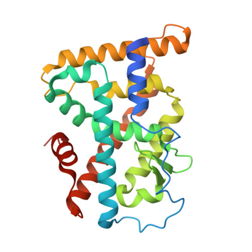
9XJZ | pdb_00009xjz

Crystal Structure of Nur77 LBD in complex with HTPA


Experimental Data Snapshot

  • Method: X-RAY DIFFRACTION
  • Resolution: 2.08 Å
  • R-Value Free: 
    0.224 (Depositor), 0.231 (DCC) 
  • R-Value Work: 
    0.180 (Depositor), 0.189 (DCC) 

Starting Model: experimental
View more details

wwPDB Validation 3D Report Full Report

Validation slider image for 9XJZ

Ligand Structure Quality Assessment 


This is version 1.0 of the entry. See complete history. 

Literature

Copper ion-induced MYH9 polymerization executes cuproptosis.

Wu, L.Z., Mi, X.Y., Liu, Z.J., Ding, Y., Hong, W.B., Cai, C.X., Hu, H.J., Ai, Y.L., Zhao, B.X., Qiu, D.D., Ma, J.S., Chen, X.Q., You, H., Lin, T., Li, F.N., Hong, X.H., Chen, H.Z., Wu, Q.

(2026) Cell 189: 6951-6967.e12

  • DOI: https://doi.org/10.1016/j.cell.2026.08.030
  • Primary Citation Related Structures: 
    9XJZ

  • PubMed Abstract: 

    Cuproptosis is a recently identified form of copper-dependent cell death implicated in various physiological and pathological processes. However, the executive proteins involved and the role of physiological intracellular copper in cuproptosis remain unclear. Herein, we identified myosin heavy-chain 9 (MYH9) as a cuproptosis executioner. Copper binding induced MYH9 polymerization, which disrupted the actin cytoskeleton and led to cuproptotic cell death. Hexyl 2-(3,4,5-trihydroxy-phenyl) acetate (HThPA) was shown to induce cuproptosis by binding to the nuclear receptor Nur77, leading to depletion of copper chelate glutathione (GSH) and release of free copper from intracellular copper stores. Acetylation of MYH9 by acetyltransferase dihydrolipoamide S-acetyltransferase (DLAT) facilitated copper binding, regardless of its extracellular or intracellular origin. Physiological copper levels were higher in drug-resistant tumor cells and clinical samples, which increased the likelihood of HThPA-induced cuproptosis. These results elucidate the mechanisms underlying cuproptosis, establish MYH9 as an executioner, and propose a treatment strategy for drug-resistant tumors.


  • Organizational Affiliation: 
    • State Key Laboratory of Cellular Stress Biology, School of Life Sciences, Xiamen University, Xiamen, Fujian 361102, China.

Macromolecule Content 

  • Total Structure Weight: 55.63 kDa 
  • Atom Count: 3,910 
  • Modeled Residue Count: 464 
  • Deposited Residue Count: 496 
  • Unique protein chains: 1

Macromolecules

Find similar proteins by:|  3D Structure
Entity ID: 1
MoleculeChains  Sequence LengthOrganismDetailsImage
Nuclear receptor subfamily 4immunitygroup A member 1
A, B
248Homo sapiensMutation(s): 0 
Gene Names: NR4A1, GFRP1, HMR, NAK1
UniProt & NIH Common Fund Data Resources
Find proteins for P22736 (Homo sapiens)
Explore P22736 
Go to UniProtKB:  P22736
GTEx:  ENSG00000123358 
Entity Groups
Sequence Clusters30% Identity50% Identity70% Identity90% Identity95% Identity100% Identity
UniProt GroupP22736
Sequence Annotations
Expand
Reference Sequence

Experimental Data & Validation

Experimental Data

  • Method: X-RAY DIFFRACTION
  • Resolution: 2.08 Å
  • R-Value Free:  0.224 (Depositor), 0.231 (DCC) 
  • R-Value Work:  0.180 (Depositor), 0.189 (DCC) 
Space Group: P 21 21 21
Unit Cell:
Length ( Å )Angle ( ˚ )
a = 74.303α = 90
b = 76.248β = 90
c = 128.597γ = 90
Software Package:
Software NamePurpose
REFMACrefinement
HKL-3000data reduction
HKL-3000data scaling
PHASERphasing

Structure Validation

View Full Validation Report


Ligand Structure Quality Assessment 


Entry History 

& Funding Information

Deposition Data


Funding OrganizationLocationGrant Number
National Natural Science Foundation of China (NSFC)China--

Revision History  (Full details and data files)

  • Version 1.0: 2026-11-11
    Type: Initial release