9XGW | pdb_00009xgw

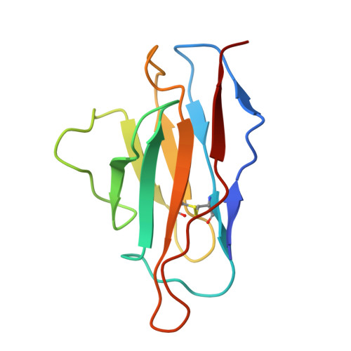
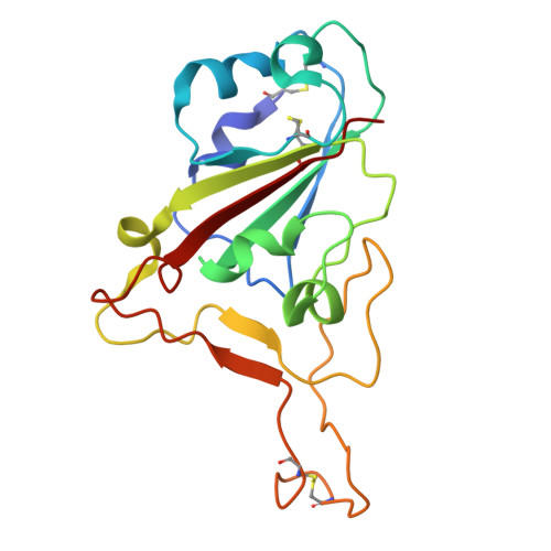
Cryo-EM structure of SARS-CoV-2 receptor binding domain in complex with CS-42 Fab (local refinement of RBD and Fv)


Experimental Data Snapshot

  • Method: ELECTRON MICROSCOPY
  • Resolution: 3.20 Å
  • Aggregation State: PARTICLE 
  • Reconstruction Method: SINGLE PARTICLE 

Starting Model: in silico
View more details

wwPDB Validation 3D Report Full Report

Validation slider image for 9XGW

This is version 1.0 of the entry. See complete history

Literature

Broad Neutralizing Activity of Monoclonal Antibodies Against the Omicron Variants Isolated From Patients With Early Severe Acute Respiratory Syndrome Coronavirus-2.

Kim, D.S.Kim, U.Woo, H.M.Lee, H.Jo, E.S.Noh, M.J.Lee, S.Y.Park, B.K.Yang, J.S.Kim, K.C.Lee, J.Y.Wang, D.M.Cho, H.S.Kim, H.J.

(2026) J Med Virol 98: e70969-e70969

  • DOI: https://doi.org/10.1002/jmv.70969
  • Primary Citation Related Structures: 
    9XGW, 9XGX

  • PubMed Abstract: 

    Since its emergence in 2019, severe acute respiratory syndrome coronavirus-2 (SARS-CoV-2) has caused a global pandemic driven by rapid mutation and high transmissibility. Current therapeutic strategies may not effectively address the continuously evolving mutant strains, underscoring the need for treatments with broad neutralizing activity. Two monoclonal antibodies (mAbs): SR-23 and CS-42, isolated from convalescent patients infected with SARS-CoV-2 during the early phase of the 2020 pandemic, demonstrated significant neutralizing activities against a variety of variants. Specifically, SR-23 exhibited neutralizing activity against BA.5 and XBB1.5, whereas CS-42 showed efficacy against Delta, BA.1, and BA.2 variants. In addition, combining the two mAbs demonstrated improved neutralization activity. Cryo-electron microscopy (cryo-EM) structural analysis revealed that SR-23 and CS-42 bind to nonoverlapping epitopes on the SARS-CoV-2 spike protein. Furthermore, both antibodies exhibited strong therapeutic effects in K18-hACE2 mice infected with D614G, BA.2, and XBB.1.5. These findings provide insights into the development of antibody-based therapeutics targeting emerging Omicron variants and suggest that combined administration could broaden neutralizing activity.


  • Organizational Affiliation
    • Division of Emerging Virus and Vector Research, Center for Emerging Virus Research, Korea National Institute of Health, Korea Disease Control and Prevention Agency, Cheongju-si, Republic of Korea.

Macromolecule Content 

  • Total Structure Weight: 46.66 kDa 
  • Atom Count: 3,257 
  • Modeled Residue Count: 416 
  • Deposited Residue Count: 420 
  • Unique protein chains: 3

Macromolecules

Find similar proteins by:|  3D Structure
Entity ID: 1
MoleculeChains  Sequence LengthOrganismDetailsImage
Heavy chain of CS-42 Fab127Homo sapiensMutation(s): 0 
Entity Groups
Sequence Clusters30% Identity50% Identity70% Identity90% Identity95% Identity100% Identity
Sequence Annotations
Expand
Reference Sequence
Find similar proteins by:|  3D Structure
Entity ID: 2
MoleculeChains  Sequence LengthOrganismDetailsImage
Light chain of CS-42 Fab109Homo sapiensMutation(s): 0 
Entity Groups
Sequence Clusters30% Identity50% Identity70% Identity90% Identity95% Identity100% Identity
Sequence Annotations
Expand
Reference Sequence
Find similar proteins by:|  3D Structure
Entity ID: 3
MoleculeChains  Sequence LengthOrganismDetailsImage
Spike protein S1C [auth E]184Severe acute respiratory syndrome coronavirus 2Mutation(s): 0 
Gene Names: S2
UniProt
Find proteins for P0DTC2 (Severe acute respiratory syndrome coronavirus 2)
Explore P0DTC2 
Go to UniProtKB:  P0DTC2
Entity Groups
Sequence Clusters30% Identity50% Identity70% Identity90% Identity95% Identity100% Identity
UniProt GroupP0DTC2
Glycosylation
Glycosylation Sites: 1Go to GlyGen: P0DTC2-1
Sequence Annotations
Expand
Reference Sequence

Small Molecules

Ligands 1 Unique
IDChains Name / Formula / InChI Key2D Diagram3D Interactions
NAG

Query on NAG


Download:Ideal Coordinates CCD File
D [auth E]2-acetamido-2-deoxy-beta-D-glucopyranose
C8 H15 N O6
OVRNDRQMDRJTHS-FMDGEEDCSA-N

Experimental Data & Validation

Experimental Data

  • Method: ELECTRON MICROSCOPY
  • Resolution: 3.20 Å
  • Aggregation State: PARTICLE 
  • Reconstruction Method: SINGLE PARTICLE 
EM Software:
TaskSoftware PackageVersion
MODEL REFINEMENTPHENIX1.20.1-4487
RECONSTRUCTIONcryoSPARC4.7.1

Structure Validation

View Full Validation Report


Entry History 

& Funding Information

Deposition Data


Funding OrganizationLocationGrant Number
National Research Foundation (NRF, Korea)Korea, Republic OfRS-2024-00344154
National Research Foundation (NRF, Korea)Korea, Republic Of2021M3A9I2080490

Revision History  (Full details and data files)

  • Version 1.0: 2026-09-09
    Type: Initial release