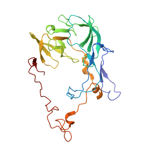
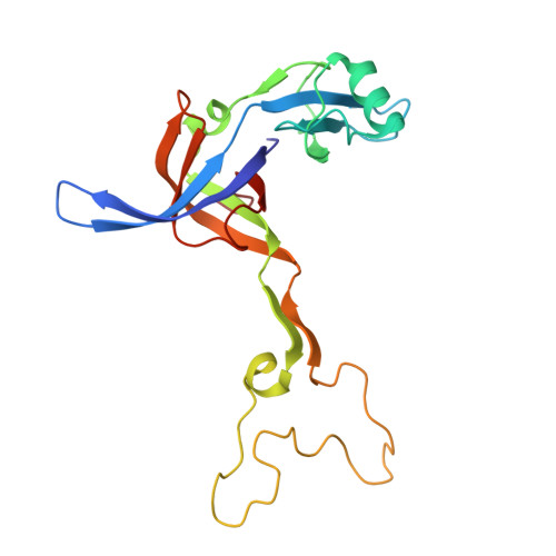
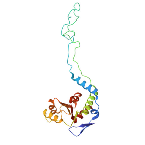
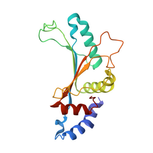
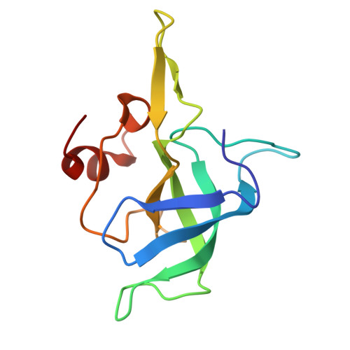
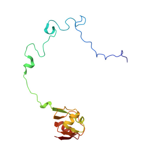
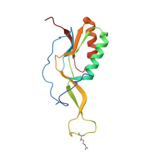
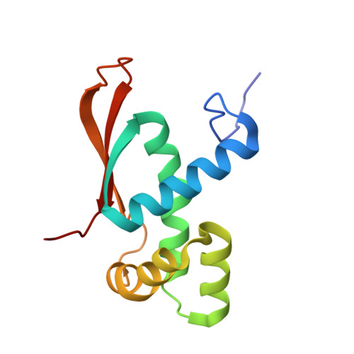
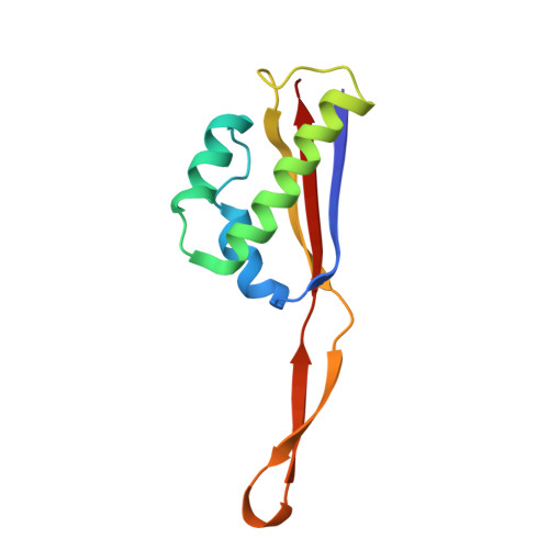
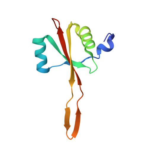
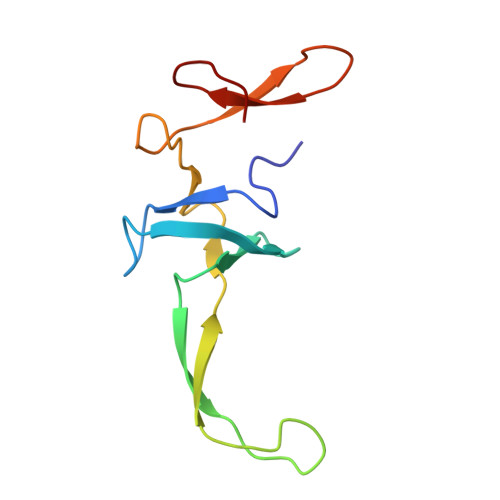
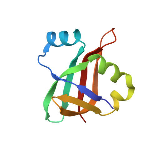
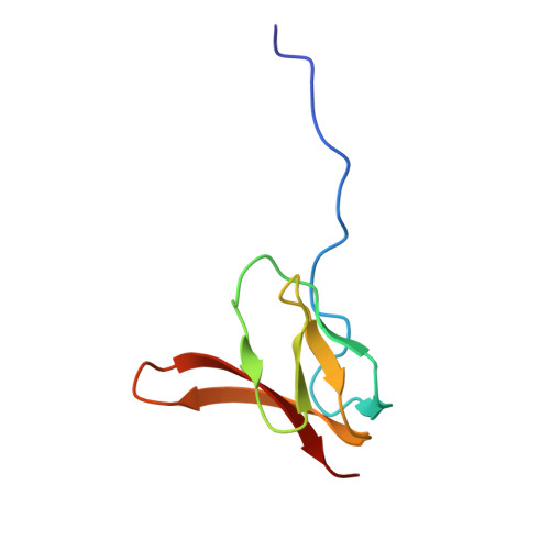
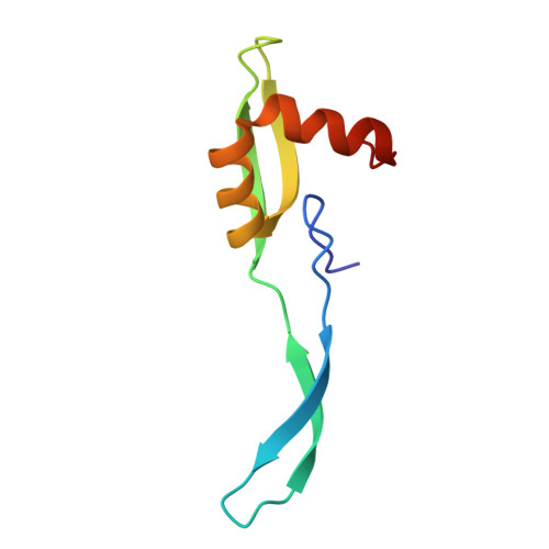
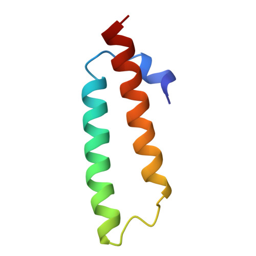
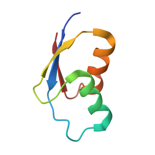
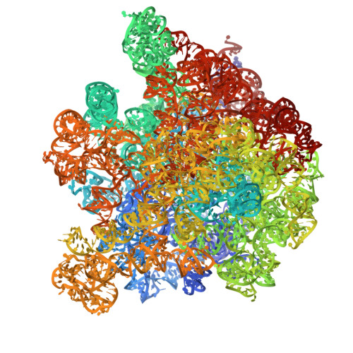
In situ structure of bacterial 50S ribosomes at 3.0 angstrom resolution from vitreous sections.
Al-Amoudi, A., Baradaran, R., Yuan, X., Wu, F., Naschberger, A.(2025) Commun Biol 9: 166-166
- PubMed: 41476251 Search on PubMedSearch on PubMed Central
- DOI: https://doi.org/10.1038/s42003-025-09441-6
- Primary Citation Related Structures:
9XFK, 9XFL - PubMed Abstract:
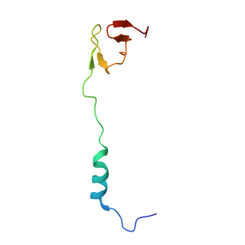
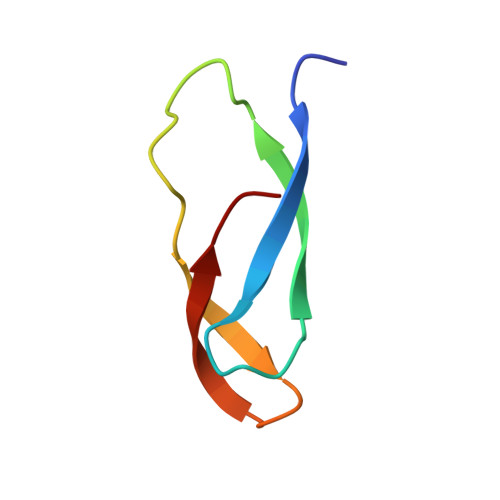
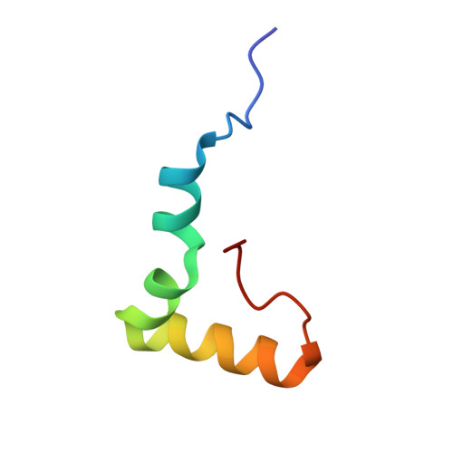
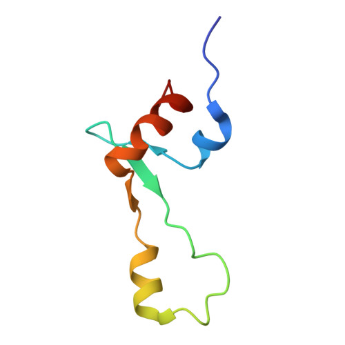
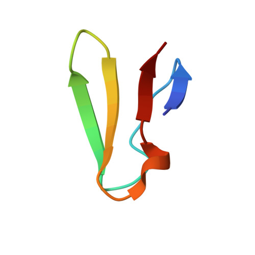
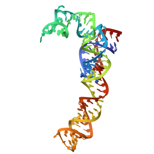
In situ high-resolution structure determination is limited to samples thin enough to be penetrated by the electron beam during imaging. Sample thinning involves focused ion or plasma beam milling of specimens to produce lamellae with thicknesses as low as 100-150 nm. However, surface damage caused by the milling process can extend 30-60 nm deep, restricting the usable lamella thickness. This imposes limitations on single-particle analysis of macromolecular complexes due to elevated structural noise, which cannot be avoided in situ because of the dense cellular environment. Alternative methods capable of producing thinner samples are needed to reduce background. Here, we demonstrate that high-resolution structures at side-chain level, free of orientation bias, can be obtained from vitreous sections prepared by cryo-ultramicrotomy, both in vitro and in situ. We optimized the method to produce sections as thin as ~40 nm, free from significant surface damage. Using this approach, we determined the structure of the 50S ribosomal subunit in vitro at 2.8 Å and in situ at 3 Å from bacterial cells. These results lay the foundation for future in situ studies of smaller complexes using CEMOVIS, as well as for methodological advances aimed at achieving compression-free sectioning.
- Electron Microscopy Lab, Imaging and Characterization Core Labs, King Abdullah University of Science and Technology (KAUST), Thuwal, Saudi Arabia. ashraf.alamoudi@kaust.edu.sa.
Organizational Affiliation: