Asymmetric sheath coordination controls flagellar architecture and function in Leptospira spirochete.
Kawamoto, A., Kuribayashi, T., Morita, M., Nakamura, S., Koizumi, N.(2026) EMBO J 45: 2882-2904
- PubMed: 41844841 Search on PubMedSearch on PubMed Central
- DOI: https://doi.org/10.1038/s44318-026-00731-1
- Primary Citation Related Structures:
9LRY, 9LRZ, 9LS0, 9LS1, 9X7K, 9X7L, 9X7M, 9X7S, 9X7V, 9X80 - PubMed Abstract:
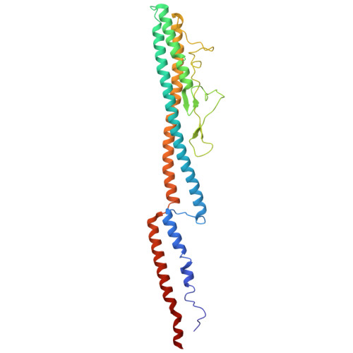
Bacterial flagella are essential for motility, but their structure and how they generate movement vary greatly. Most motile bacteria use external helical flagella, whereas spirochetes have periplasmic flagella (PFs) that distort the cell body to drive forward movement. Here, we generated sheath protein knockout mutants and used high-resolution cryo-electron microscopy to elucidate the mechanisms underlying PF assembly, curvature, and rigidity in Leptospira biflexa. The PF consists of a FlaB1-based core filament surrounded asymmetrically by sheath proteins. Weak but essential binding of FlaA2 to the core enables asymmetric localization of the coiling protein FcpA. FcpA alone can induce curvature, whereas FcpB acts as a structural wedge that reinforces PF rigidity and enables efficient swimming in liquid. Specific glycosylation of FlaB1 mediates sheath-core interactions and may guide the assembly of sheath components. We propose that sheath proteins interact transiently with the core and may be anchored to the outer membrane, allowing core rotation beneath a static sheath. These findings reveal how cooperative interactions among sheath components confer structural and mechanical specialization to spirochete flagella.
- Institute for Protein Research, The University of Osaka, 3-2 Yamadaoka, Suita, Osaka, 565-0871, Japan. kawamoto@protein.osaka-u.ac.jp.
Organizational Affiliation: