9X0I | pdb_00009x0i

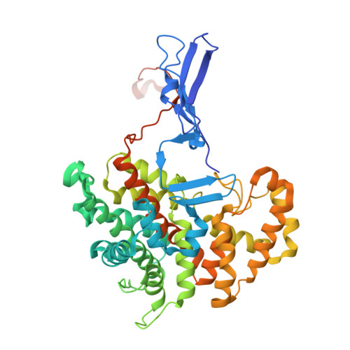
Glyoxysomal Citrate Synthase 3 from Arabidopsis thaliana in complex with OAA and CoA


Experimental Data Snapshot

  • Method: X-RAY DIFFRACTION
  • Resolution: 1.70 Å
  • R-Value Free: 
    0.197 (Depositor), 0.206 (DCC) 
  • R-Value Work: 
    0.166 (Depositor), 0.172 (DCC) 

Starting Model: experimental
View more details

wwPDB Validation 3D Report Full Report

Validation slider image for 9X0I

Ligand Structure Quality Assessment 


This is version 1.1 of the entry. See complete history

Literature

Crystal structure of glyoxysomal citrate synthase 3 from Arabidopsis thaliana reveals a novel oligomeric state.

Nishio, K.Takagi, K.Mizushima, T.

(2026) J Struct Biol 218: 108293-108293

  • DOI: https://doi.org/10.1016/j.jsb.2026.108293
  • Primary Citation Related Structures: 
    9X0I, 9XAU, 9XDG

  • PubMed Abstract: 

    Citrate synthase (CS) is a pivotal enzyme in carbohydrate and energy metabolism, with distinct isoforms present in various eukaryotic compartments, including mitochondria and glyoxysomes in plants. While CSs exhibit diverse oligomeric states, detailed structural information on higher plant non-mitochondrial Type II CSs has been limited. We herein determined the crystal structures of CS 3 from Arabidopsis thaliana (AtCSY3) in complex with oxaloacetate (OAA) and acetyl-coenzyme A (CoA)-OAA at resolutions of 2.0 and 1.7 Å, respectively. These structures revealed that AtCSY3 can form a homo-tetrameric assembly that is distinct from the hexameric Escherichia coli CS and the octameric Ananas comosus CS. The tetrameric arrangement observed in the crystal structure is mediated by hydrogen-bonding and hydrophobic interactions between subunits. Gel filtration chromatography further suggests the presence of a tetrameric species in solution under the purification conditions. Ligand density was observed near the interface between the two dimers in the tetrameric structure; however, no experimental evidence is currently available to determine whether ligand binding affects the oligomeric state or enzymatic activity of AtCSY3. These structures illustrate the structural diversity of CS oligomerization and provide a structural basis for studies of plant glyoxysomal CSs.


  • Organizational Affiliation
    • Department of Science, Graduate School of Science, University of Hyogo, 2167, Shosha, Himeji 671-2280, Japan.

Macromolecule Content 

  • Total Structure Weight: 108 kDa 
  • Atom Count: 7,902 
  • Modeled Residue Count: 881 
  • Deposited Residue Count: 958 
  • Unique protein chains: 1

Macromolecules

Find similar proteins by:|  3D Structure
Entity ID: 1
MoleculeChains  Sequence LengthOrganismDetailsImage
Citrate synthase 3, peroxisomal
A, B
479Arabidopsis thalianaMutation(s): 0 
Gene Names: CSY3At2g42790F7D19.21
EC: 2.3.3.16
UniProt
Find proteins for Q9SJH7 (Arabidopsis thaliana)
Explore Q9SJH7 
Go to UniProtKB:  Q9SJH7
Entity Groups
Sequence Clusters30% Identity50% Identity70% Identity90% Identity95% Identity100% Identity
UniProt GroupQ9SJH7
Sequence Annotations
Expand
Reference Sequence

Small Molecules

Ligands 6 Unique
IDChains Name / Formula / InChI Key2D Diagram3D Interactions
COA

Query on COA


Download:Ideal Coordinates CCD File
D [auth A],
K [auth B]
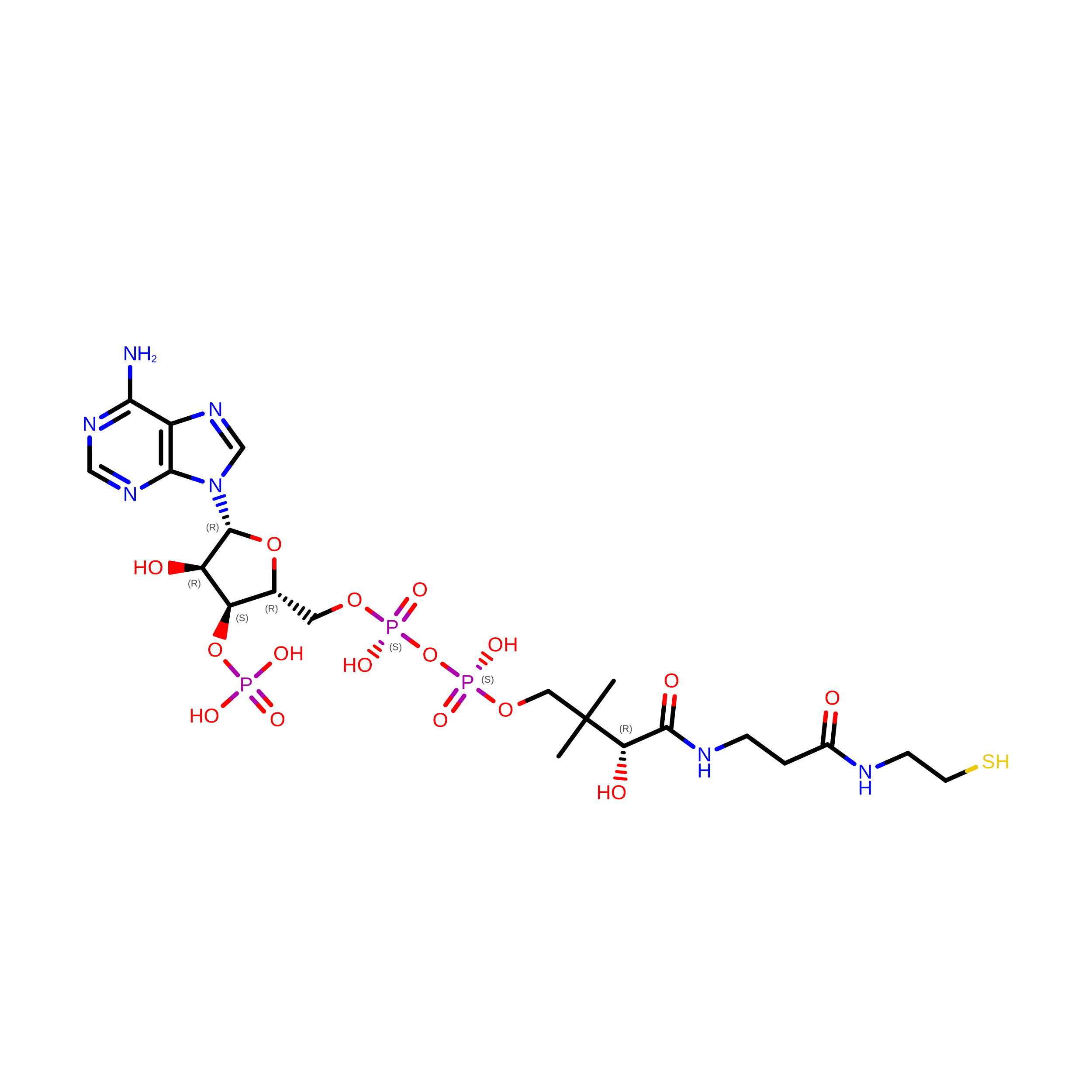
COENZYME A
C21 H36 N7 O16 P3 S
RGJOEKWQDUBAIZ-IBOSZNHHSA-N
EPE

Query on EPE


Download:Ideal Coordinates CCD File
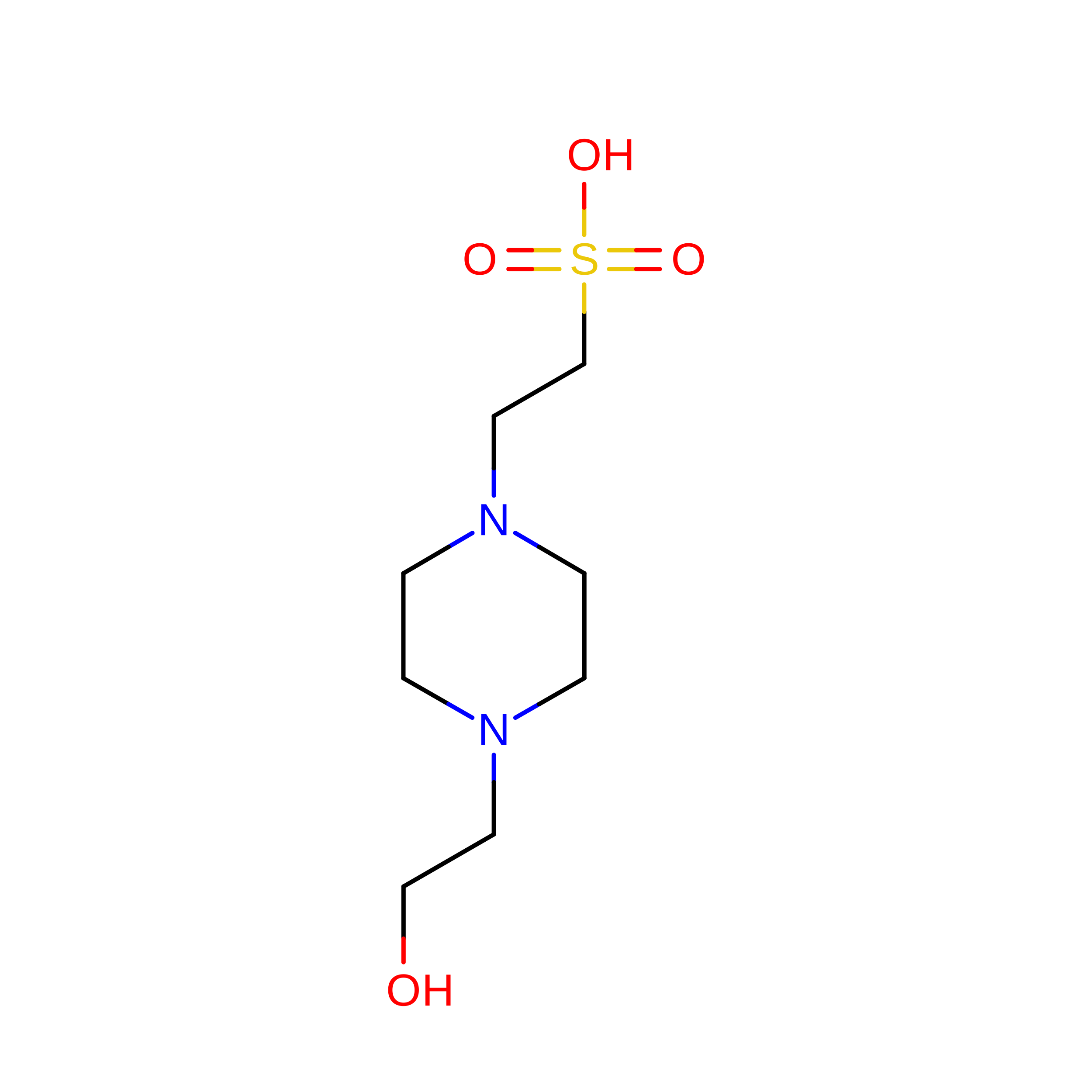
O [auth B]4-(2-HYDROXYETHYL)-1-PIPERAZINE ETHANESULFONIC ACID
C8 H18 N2 O4 S
JKMHFZQWWAIEOD-UHFFFAOYSA-N
OAA

Query on OAA


Download:Ideal Coordinates CCD File
C [auth A],
J [auth B]
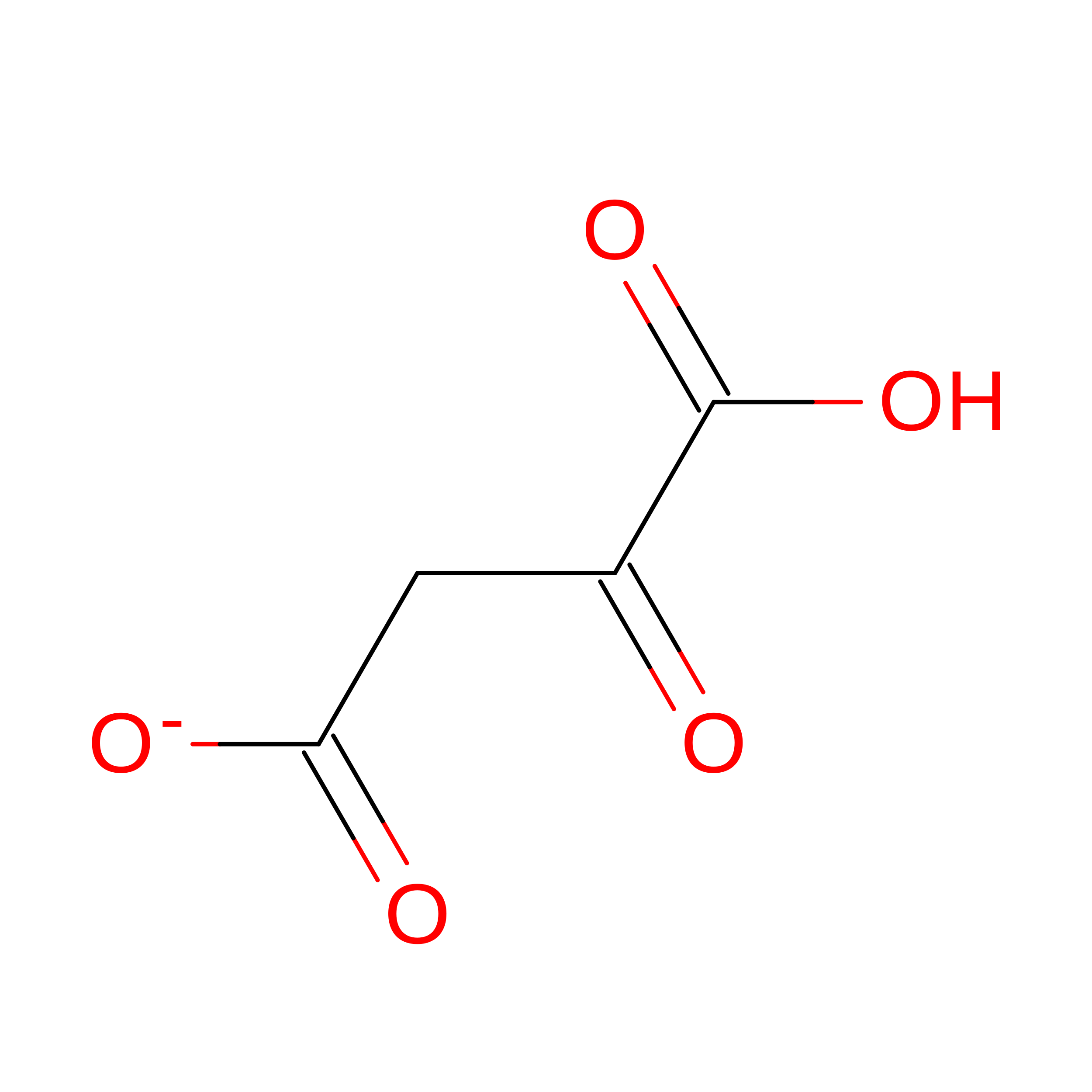
OXALOACETATE ION
C4 H3 O5
KHPXUQMNIQBQEV-UHFFFAOYSA-M
PEG

Query on PEG


Download:Ideal Coordinates CCD File
E [auth A],
F [auth A]
DI(HYDROXYETHYL)ETHER
C4 H10 O3
MTHSVFCYNBDYFN-UHFFFAOYSA-N
EDO

Query on EDO


Download:Ideal Coordinates CCD File
L [auth B]1,2-ETHANEDIOL
C2 H6 O2
LYCAIKOWRPUZTN-UHFFFAOYSA-N
MG

Query on MG


Download:Ideal Coordinates CCD File
G [auth A],
H [auth A],
I [auth A],
M [auth B],
N [auth B]
MAGNESIUM ION
Mg
JLVVSXFLKOJNIY-UHFFFAOYSA-N

Experimental Data & Validation

Experimental Data

  • Method: X-RAY DIFFRACTION
  • Resolution: 1.70 Å
  • R-Value Free:  0.197 (Depositor), 0.206 (DCC) 
  • R-Value Work:  0.166 (Depositor), 0.172 (DCC) 
Space Group: P 21 21 2
Unit Cell:
Length ( Å )Angle ( ˚ )
a = 193.859α = 90
b = 60.597β = 90
c = 76.864γ = 90
Software Package:
Software NamePurpose
REFMACrefinement
XDSdata reduction
XDSdata scaling
PHASERphasing

Structure Validation

View Full Validation Report


Ligand Structure Quality Assessment 


Entry History 

& Funding Information

Deposition Data


Funding OrganizationLocationGrant Number
Japan Society for the Promotion of Science (JSPS)Japan24K01968

Revision History  (Full details and data files)

  • Version 1.0: 2026-02-04
    Type: Initial release
  • Version 1.1: 2026-02-18
    Changes: Database references