9WRN | pdb_00009wrn

Crystal structure of chimeric anti-Z-DNA Fab cZ22-Fab


Experimental Data Snapshot

  • Method: X-RAY DIFFRACTION
  • Resolution: 2.15 Å
  • R-Value Free: 
    0.238 (Depositor), 0.238 (DCC) 
  • R-Value Work: 
    0.192 (Depositor), 0.192 (DCC) 
  • R-Value Observed: 
    0.195 (Depositor) 

Starting Model: experimental
View more details

wwPDB Validation   3D Report Full Report


This is version 1.1 of the entry. See complete history


Literature

Structural basis of B-to-Z DNA transition mediated by an anti-Z-DNA antibody.

Lee, C.C.Hsu, S.F.Chang, Y.W.Chen, Y.W.Ho, M.R.Sugiyama, H.Wang, W.C.Wang, A.H.

(2026) Nucleic Acids Res 54

  • DOI: https://doi.org/10.1093/nar/gkag160
  • Primary Citation of Related Structures:  
    9WRN, 9WS0, 9WU2

  • PubMed Abstract: 

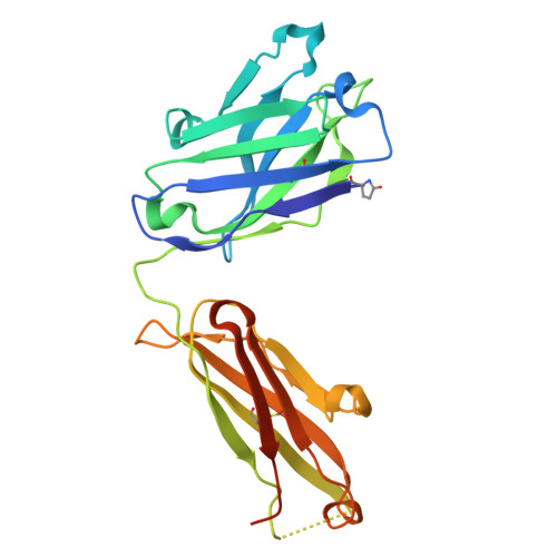
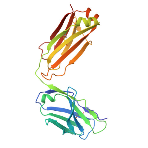
    Z-DNA is a left-handed double-helical form of DNA that plays roles in transcription, immune responses, viral infection, bacterial biofilm formation, and autoimmune diseases. Despite its importance, the instability of Z-DNA under physiological conditions has hindered detailed structural and functional investigations. Moreover, although antibodies are known to recognize nucleic acids, the mechanisms underlying their detection and stabilization of dynamic DNA under biological conditions remain unclear. This study provides the first atomic-level structural insights into antibody-mediated B-to-Z DNA transition. Accordingly, a Z-DNA-specific chimeric Fab fragment of Z22 (cZ22-Fab) was designed and characterized using multiple biophysical approaches. cZ22-Fab mediates a concentration-dependent B-to-Z conformational transition in CG-repeat DNA, establishing a stable 2:1 Fab/DNA stoichiometry. Crystal structure of cZ22-Fab/Z-DNA complexes revealed a left-handed DNA backbone-tracking recognition mode, in which cZ22-Fab recognizes Z-DNA conformation through phosphate-clamping and base interactions. Notably, a 5'-end C-hanging Z-DNA duplex structure formed by dC(GC)3 and stabilized by cZ22-Fab was observed. Structure-guided mutagenesis demonstrated that heavy chain residues R50 and Y106 are critical for Z-DNA binding, and analyses of additional Z-DNA-forming sequences further elucidated the binding characteristics. Overall, this work provides molecular insights into the mechanism of antibody-mediated Z-DNA formation and stabilization, highlighting its therapeutic relevance and implications for autoimmunity.


  • Organizational Affiliation
    • The Ph.D. Program for Translational Medicine, College of Medical Science and Technology, Taipei Medical University, Taipei 110301, Taiwan.

Macromolecules
Find similar proteins by:  (by identity cutoff)  |  3D Structure
Entity ID: 1
MoleculeChains Sequence LengthOrganismDetailsImage
cZ22-Fab heavy chainA [auth H],
B [auth I],
C [auth J],
D [auth K]
236Mus musculusMutation(s): 0 
Entity Groups  
Sequence Clusters30% Identity50% Identity70% Identity90% Identity95% Identity100% Identity
Sequence Annotations
Expand
  • Reference Sequence
Find similar proteins by:  (by identity cutoff)  |  3D Structure
Entity ID: 2
MoleculeChains Sequence LengthOrganismDetailsImage
cZ22-Fab light chainE [auth L],
F [auth M],
G [auth N],
H [auth O]
214Mus musculusMutation(s): 0 
Entity Groups  
Sequence Clusters30% Identity50% Identity70% Identity90% Identity95% Identity100% Identity
Sequence Annotations
Expand
  • Reference Sequence
Small Molecules
Modified Residues  1 Unique
IDChains TypeFormula2D DiagramParent
PCA
Query on PCA
A [auth H],
B [auth I],
C [auth J],
D [auth K]
L-PEPTIDE LINKINGC5 H7 N O3GLN
Experimental Data & Validation

Experimental Data

  • Method: X-RAY DIFFRACTION
  • Resolution: 2.15 Å
  • R-Value Free:  0.238 (Depositor), 0.238 (DCC) 
  • R-Value Work:  0.192 (Depositor), 0.192 (DCC) 
  • R-Value Observed: 0.195 (Depositor) 
Space Group: P 1 21 1
Unit Cell:
Length ( Å )Angle ( ˚ )
a = 74.016α = 90
b = 87.025β = 95.47
c = 149.4γ = 90
Software Package:
Software NamePurpose
PHENIXrefinement
XDSdata reduction
XDSdata scaling
PHENIXphasing

Structure Validation

View Full Validation Report


Entry History & Funding Information

Deposition Data


Funding OrganizationLocationGrant Number
Academia Sinica (Taiwan)Taiwan--

Revision History  (Full details and data files)

  • Version 1.0: 2026-02-18
    Type: Initial release
  • Version 1.1: 2026-03-18
    Changes: Database references