Structural basis for spectral red shift and UVA absorption in the microalgal water-soluble astaxanthin-binding protein AstaP-pink1.
Mitsui, T., Shomura, Y., Furubayashi, M., Kato, R., Takaichi, S., Kawasaki, S.(2026) J Struct Biol 218: 108288-108288
- PubMed: 41500483 Search on PubMed
- DOI: https://doi.org/10.1016/j.jsb.2026.108288
- Primary Citation Related Structures:
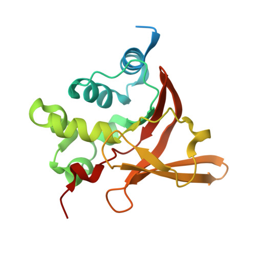
9WM8 - PubMed Abstract:
AstaPs are water-soluble, photooxidative stress-inducible astaxanthin (AXT)-binding proteins found only in Scenedesmaceae microalgae, where they play a central role in survival under severe photooxidative stress. Here, we focused on the unique function of AstaP-pink1, which converts orange AXT into a pink form and generates a UVA absorption spectrum upon protein binding. AstaP-pink1 was expressed in genetically engineered Escherichia coli strains capable of synthesizing AXT. The host strain harboring pAC-Asta produced adonixanthin, AXT, and zeaxanthin in an approximate ratio of 5:3:2, whereas the strain carrying pMF573 predominantly produced AXT (∼90 % of total carotenoid). Co-expression of the gene encoding AstaP-pink1 in these strains resulted in moderate and selective AXT binding, accompanied by a spectral red shift and UVA absorption, thereby generating pink coloration. Crystal structure analysis of AXT-bound recombinant AstaP-pink1 (rAstaP-pink1) revealed both similarities and differences in AXT binding compared with rAstaP-orange1. Density functional theory (DFT) calculations based on the crystal structure suggested that the larger red shift than that of AstaP-orange1 and the distinct UVA absorption are derived from the conformation of AXT that is compelled by binding to AstaP-pink1. This study suggests that AXT binding by AstaP-pink1 not only facilitates the water solubilization of AXT but also generates the observed spectral properties.
- Department of Molecular Microbiology, Tokyo University of Agriculture, 1-1-1 Sakuragaoka, Setagaya-ku, Tokyo 156-8502, Japan.
Organizational Affiliation: