Download Mendeley
Structural basis of Saccharomyces cerevisiae Mba1 in mitochondrial co-translational membrane insertion.
Yang, J., Ruan, M., Bai, D., Liu, Y., Wang, Y., Tang, H., Li, Y., Wang, J.(2026) J Struct Biol 218: 108359-108359
- PubMed: 42660304 Search on PubMed
- DOI: https://doi.org/10.1016/j.jsb.2026.108359
- Primary Citation Related Structures:
9WKT - PubMed Abstract:
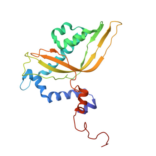
Co-translational membrane insertion is essential for the efficient integration of mitochondrially encoded proteins into the inner mitochondrial membrane (IMM) and is critical for respiratory chain biogenesis. Mba1 is a mitochondrial ribosome-associated protein implicated in coupling mitochondrial translation with inner-membrane protein biogenesis, but its structural basis of function remains poorly understood. Here, we determined the solution structure of mature Saccharomyces cerevisiae Mba1 (mMba1) using multidimensional nuclear magnetic resonance (NMR) spectroscopy. The structure reveals a compact α + β fold with a central hydrophobic cavity and distinct charged surface regions. Ribosome titration, paramagnetic relaxation enhancement, and Cox2-derived peptide titration identified several regions of mMba1 that are affected by these different interaction conditions. Mapping these regions onto the structure reveals spatially distinct surfaces that may contribute to ribosome association, membrane proximity, and interactions with hydrophobic peptide segments. These findings provide a structural framework for interpreting previous functional studies of Mba1 and support a working model in which Mba1 may function as a peripheral adaptor at the mitoribosome-inner membrane interface. Further structural and biochemical studies will be required to establish the molecular mechanisms underlying these interactions.
- High Magnetic Field Laboratory, Key Laboratory of High Magnetic Field and Ion Beam Physical Biology, Hefei Institutes of Physical Science, Chinese Academy of Sciences, Hefei, China.
Organizational Affiliation: