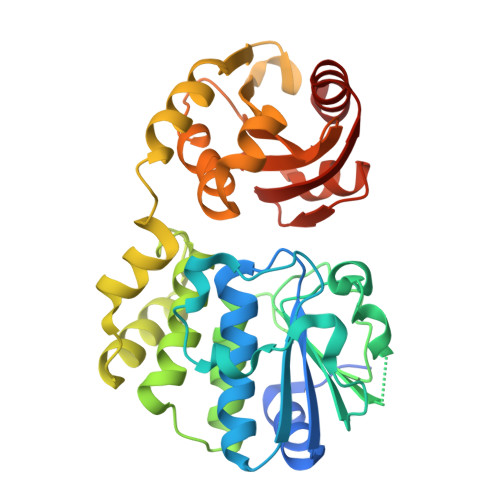
9WBU | pdb_00009wbu

Crystal structure of Mtb cyclic dinucleotide phosphodiesterase by sulfur-modified cyclic dinucleotide analogue


Experimental Data Snapshot

  • Method: X-RAY DIFFRACTION
  • Resolution: 2.96 Å
  • R-Value Free: 
    0.270 (Depositor), 0.275 (DCC) 
  • R-Value Work: 
    0.218 (Depositor), 0.222 (DCC) 
  • R-Value Observed: 
    0.221 (Depositor) 

Starting Model: experimental
View more details

wwPDB Validation 3D Report Full Report

Validation slider image for 9WBU

Ligand Structure Quality Assessment 


This is version 1.1 of the entry. See complete history. 

Literature

Structural and biochemical insights into the inhibition of Mycobacterium tuberculosis cyclic dinucleotide phosphodiesterase by a sulfur-modified cyclic dinucleotide analog.

Hanuman, D.S., Neeharika, S., Murari, S.K., Yeboah, S.K., Sintim, H.O., Rajakumara, E.

(2026) RSC Chem Biol 7: 880-891

  • DOI: https://doi.org/10.1039/d6cb00006a
  • Primary Citation Related Structures: 
    9WBU

  • PubMed Abstract: 

    Cyclic dinucleotide (CDN) signaling plays a pivotal role in bacterial physiology and host-pathogen interactions. Mycobacterium tuberculosis (Mtb) releases a cyclic dinucleotide phosphodiesterase (CdnP) in the macrophages, which hydrolyzes Mtb-derived 3'3'-c-di-AMP and 3'3'-c-di-GMP, and a host-derived 2'3'-cGAMP STING agonist, to evade the host's innate immune response mediated by the STING protein. Therefore, by inhibiting CdnP released into host cells, the STING pathway can be potentiated, leading to improved bacterial clearance, which represents a potential novel approach for anti-tuberculosis (anti-TB) therapy. Here we report ES-2'3'-cAAMP, an analog of the host-derived STING agonist 2'3'-cGAMP, in which the phosphodiester bond is modified and the base is replaced, binds to CdnP with a micromolar binding affinity and competes with CdnP's substrates - 3'3'-c-di-AMP and 3'3'-c-di-GMP - binding to CdnP. Significantly, CdnP's phosphodiesterase catalytic activity is inhibited by ES-2'3'-cAAMP. Furthermore, the CdnP-ES-2'3'-cAAMP complex structure reported here is the first structure of the CdnP complexed with CDN, revealing the unique pose of ES-2'3'-cAAMP in the catalytic pocket of CdnP that is inaccessible to catalytic residues and Mn 2+ ions for its hydrolysis, and in parallel blocks the binding of the natural substrates of CdnP that explains the structural basis of CdnP's catalytic activity inhibition by the inhibitor. Additionally, 2'3'-cGAMP in the STING receptor and ES-2'3'-cAAMP in the CdnP adopted an identical horseshoe conformation, suggesting that ES-2'3'-cAAMP, or an analogue thereof, can bind to and stimulate STING, thus acting as a synthetic STING agonist. These combined structural and biochemical findings provide new mechanistic insights into the inhibition of Mtb CdnP and offer a novel approach to host-directed anti-TB therapies that aim to enhance the host's own immune responses rather than directly killing the pathogen, which may help to mitigate the problem of antibiotic resistance.


  • Organizational Affiliation: 
    • Macromolecular Structural Biology Laboratory, Department of Biotechnology, Indian Institute of Technology Hyderabad (IITH) Hyderabad Telangana-502285 India eraj@bt.iith.ac.in.

Macromolecule Content 

  • Total Structure Weight: 108.62 kDa 
  • Atom Count: 6,912 
  • Modeled Residue Count: 940 
  • Deposited Residue Count: 1,008 
  • Unique protein chains: 1

Macromolecules

Find similar proteins by:|  3D Structure
Entity ID: 1
MoleculeChains  Sequence LengthOrganismDetailsImage
Bifunctional oligoribonuclease and PAP phosphatase NrnAA [auth B],
B [auth C],
C [auth A]
336Mycobacterium tuberculosis H37RvMutation(s): 0 
Gene Names: nrnA, Rv2837c
EC: 3.1 (PDB Primary Data), 3.1.3.7 (PDB Primary Data)
UniProt
Find proteins for P71615 (Mycobacterium tuberculosis (strain ATCC 25618 / H37Rv))
Explore P71615 
Go to UniProtKB:  P71615
Entity Groups
Sequence Clusters30% Identity50% Identity70% Identity90% Identity95% Identity100% Identity
UniProt GroupP71615
Sequence Annotations
Expand
Reference Sequence

Small Molecules

Ligands 3 Unique
IDChains Name / Formula / InChI Key2D Diagram3D Interactions
A1MBE
(Subject of Investigation/LOI)

Query on A1MBE


Download:Ideal Coordinates CCD File
D [auth B],
G [auth C],
I [auth A]
(1~{R},6~{S},8~{R},9~{R},10~{S},15~{R},17~{R},18~{R})-8,17-bis(6-aminopurin-9-yl)-3,12-bis(oxidanyl)-3,12-bis(oxidanylidene)-2,7,11,13,16-pentaoxa-4-thia-3$l^{5},12$l^{5}-diphosphatricyclo[13.2.1.0^{6,10}]octadecane-9,18-diol
C20 H24 N10 O11 P2 S
FKYKADUIHLPICL-XPWFQUROSA-N
EDO

Query on EDO


Download:Ideal Coordinates CCD File
E [auth B]1,2-ETHANEDIOL
C2 H6 O2
LYCAIKOWRPUZTN-UHFFFAOYSA-N
MN

Query on MN


Download:Ideal Coordinates CCD File
F [auth B],
H [auth C],
J [auth A]
MANGANESE (II) ION
Mn
WAEMQWOKJMHJLA-UHFFFAOYSA-N

Experimental Data & Validation

Experimental Data

  • Method: X-RAY DIFFRACTION
  • Resolution: 2.96 Å
  • R-Value Free:  0.270 (Depositor), 0.275 (DCC) 
  • R-Value Work:  0.218 (Depositor), 0.222 (DCC) 
  • R-Value Observed: 0.221 (Depositor) 
Space Group: I 21 21 21
Unit Cell:
Length ( Å )Angle ( ˚ )
a = 79.01α = 90
b = 149.096β = 90
c = 167.033γ = 90
Software Package:
Software NamePurpose
REFMACrefinement
Aimlessdata scaling
MOLREPphasing
PDB_EXTRACTdata extraction
iMOSFLMdata reduction

Structure Validation

View Full Validation Report


Ligand Structure Quality Assessment 


Entry History 

& Funding Information

Deposition Data


Funding OrganizationLocationGrant Number
Science and Engineering Research Board (SERB)IndiaCRG/2020/003946

Revision History  (Full details and data files)

  • Version 1.0: 2026-08-26
    Type: Initial release
  • Version 1.1: 2026-09-16
    Changes: Database references, Structure summary