9W74 | pdb_00009w74

Cryo-EM structure of the close-packed di-hexasome (CPDH)


Experimental Data Snapshot

  • Method: ELECTRON MICROSCOPY
  • Resolution: 4.94 Å
  • Aggregation State: PARTICLE 
  • Reconstruction Method: SINGLE PARTICLE 

wwPDB Validation   3D Report Full Report


This is version 1.1 of the entry. See complete history


Literature

A method for cryo-EM analysis of eukaryotic nucleosomes reconstituted in bacterial cells.

Ho, C.H.Kobayashi, Y.Ogasawara, M.Takizawa, Y.Kurumizaka, H.

(2026) iScience 29: 114453-114453

  • DOI: https://doi.org/10.1016/j.isci.2025.114453
  • Primary Citation of Related Structures:  
    9W74

  • PubMed Abstract: 

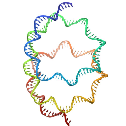
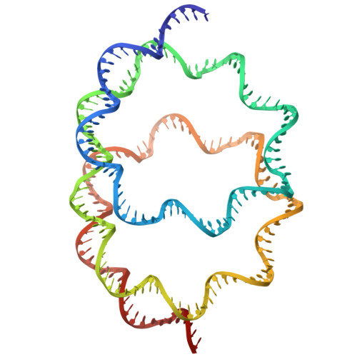
    Conventional methods for preparing nucleosomes are time-consuming and technically demanding. In the present study, we extended the approach of generating nucleosomes in Escherichia coli by the co-expression of all four histones, allowing nucleosomes to be assembled in cells. The bacterially reconstituted nucleosomes can be readily prepared from the E. coli cells and directly subjected to cryo-EM single particle analysis. Using this method, we obtained a 2.56 Å nucleosome structure that is highly similar to a previously reported nucleosome crystal structure, validating the use of nucleosomes formed in E. coli for cryo-EM analysis. Unexpectedly, we also discovered a non-canonical nucleosome structure, in which two hexasomes are closely packed. This system provides a robust and efficient platform for structural studies of nucleosomes and nucleosome variants, and may facilitate the discovery of chromatin architectures.


  • Organizational Affiliation
    • Laboratory of Chromatin Structure and Function, Institute for Quantitative Biosciences, The University of Tokyo, 1-1-1 Yayoi, Bunkyo-ku, Tokyo 113-0032, Japan.

Macromolecules

Find similar proteins by:  (by identity cutoff)  |  3D Structure
Entity ID: 1
MoleculeChains Sequence LengthOrganismDetailsImage
Histone H3.2A,
E,
G [auth K],
K [auth O]
136Xenopus laevisMutation(s): 1 
UniProt
Find proteins for P84233 (Xenopus laevis)
Explore P84233 
Go to UniProtKB:  P84233
Entity Groups  
Sequence Clusters30% Identity50% Identity70% Identity90% Identity95% Identity100% Identity
UniProt GroupP84233
Sequence Annotations
Expand
  • Reference Sequence
Find similar proteins by:  (by identity cutoff)  |  3D Structure
Entity ID: 2
MoleculeChains Sequence LengthOrganismDetailsImage
Histone H4B,
F,
H [auth L],
L [auth P]
120Xenopus laevisMutation(s): 0 
UniProt
Find proteins for P62799 (Xenopus laevis)
Explore P62799 
Go to UniProtKB:  P62799
Entity Groups  
Sequence Clusters30% Identity50% Identity70% Identity90% Identity95% Identity100% Identity
UniProt GroupP62799
Sequence Annotations
Expand
  • Reference Sequence
Find similar proteins by:  (by identity cutoff)  |  3D Structure
Entity ID: 3
MoleculeChains Sequence LengthOrganismDetailsImage
Histone H2A type 1C,
I [auth M]
165Xenopus laevisMutation(s): 2 
UniProt
Find proteins for P06897 (Xenopus laevis)
Explore P06897 
Go to UniProtKB:  P06897
Entity Groups  
Sequence Clusters30% Identity50% Identity70% Identity90% Identity95% Identity100% Identity
UniProt GroupP06897
Sequence Annotations
Expand
  • Reference Sequence
Find similar proteins by:  (by identity cutoff)  |  3D Structure
Entity ID: 4
MoleculeChains Sequence LengthOrganismDetailsImage
Histone H2B 1.1D,
J [auth N]
123Xenopus laevisMutation(s): 1 
UniProt
Find proteins for P02281 (Xenopus laevis)
Explore P02281 
Go to UniProtKB:  P02281
Entity Groups  
Sequence Clusters30% Identity50% Identity70% Identity90% Identity95% Identity100% Identity
UniProt GroupP02281
Sequence Annotations
Expand
  • Reference Sequence
Find similar nucleic acids by:  (by identity cutoff)  |  3D Structure
Entity ID: 5
MoleculeChains LengthOrganismImage
DNA (207-MER)M [auth I]207Escherichia coli
Sequence Annotations
Expand
  • Reference Sequence
Find similar nucleic acids by:  (by identity cutoff)  |  3D Structure
Entity ID: 6
MoleculeChains LengthOrganismImage
DNA (207-MER)N [auth J]207Escherichia coli
Sequence Annotations
Expand
  • Reference Sequence
Experimental Data & Validation

Experimental Data

  • Method: ELECTRON MICROSCOPY
  • Resolution: 4.94 Å
  • Aggregation State: PARTICLE 
  • Reconstruction Method: SINGLE PARTICLE 
EM Software:
TaskSoftware PackageVersion
RECONSTRUCTIONcryoSPARC4.7.1

Structure Validation

View Full Validation Report


Entry History & Funding Information

Deposition Data


Funding OrganizationLocationGrant Number
Japan Society for the Promotion of Science (JSPS)JapanJP23H05475
Japan Society for the Promotion of Science (JSPS)JapanJP24H02319
Japan Society for the Promotion of Science (JSPS)JapanJP24H02328
Japan Science and TechnologyJapanJPMJER1901
Japan Science and TechnologyJapanJPMJCR24T3
Japan Agency for Medical Research and Development (AMED)JapanJP25ama121009
Japan Agency for Medical Research and Development (AMED)JapanJP25ama121002
Japan Society for the Promotion of Science (JSPS)JapanJP25K18403

Revision History  (Full details and data files)

  • Version 1.0: 2025-12-10
    Type: Initial release
  • Version 1.1: 2026-01-28
    Changes: Data collection, Database references